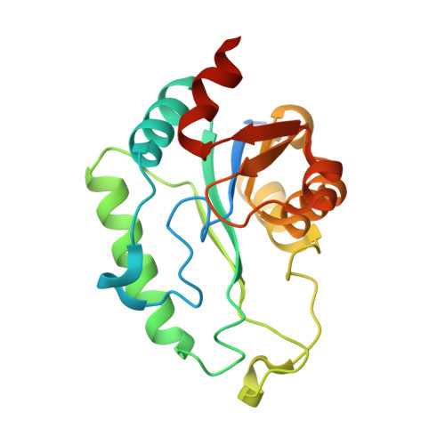
Structural and kinetic characterization of the LPS biosynthetic enzyme D-alpha,beta-D-heptose-1,7-bisphosphate phosphatase (GmhB) from Escherichia coli.
Taylor, P.L., Sugiman-Marangos, S., Zhang, K., Valvano, M.A., Wright, G.D., Junop, M.S.(2010) Biochemistry 49: 1033-1041
- PubMed: 20050699 Search on PubMed
- DOI: https://doi.org/10.1021/bi901780j
- Primary Citation Related Structures:
2GMW, 3L1U, 3L1V - PubMed Abstract:
Lipopolysaccharide is a major component of the outer membrane of gram-negative bacteria and provides a permeability barrier to many commonly used antibiotics. ADP-heptose residues are an integral part of the LPS inner core, and mutants deficient in heptose biosynthesis demonstrate increased membrane permeability. The heptose biosynthesis pathway involves phosphorylation and dephosphorylation steps not found in other pathways for the synthesis of nucleotide sugar precursors. Consequently, the heptose biosynthetic pathway has been marked as a novel target for antibiotic adjuvants, which are compounds that facilitate and potentiate antibiotic activity. D-alpha,beta-D-heptose-1,7-bisphosphate phosphatase (GmhB) catalyzes the third essential step of LPS heptose biosynthesis. This study describes the first crystal structure of GmhB and enzymatic analysis of the protein. Structure-guided mutations followed by steady state kinetic analysis, together with established precedent for HAD phosphatases, suggest that GmhB functions through a phosphoaspartate intermediate. This study provides insight into the structure-function relationship of GmhB, a new target for combatting gram-negative bacterial infection.
- Department of Biochemistry and Biomedical Sciences and M. G. DeGroote Institute for Infectious Disease Research, McMaster University, 1200 Main Street West, Hamilton, Ontario L8N 3Z5, Canada.
Organizational Affiliation: