3KX1 | pdb_00003kx1

Cathepsin K in complex with a selective 2-cyano-pyrimidine inhibitor


Experimental Data Snapshot

  • Method: X-RAY DIFFRACTION
  • Resolution: 1.51 Å
  • R-Value Free: 
    0.306 (Depositor), 0.302 (DCC) 
  • R-Value Work: 
    0.239 (Depositor), 0.239 (DCC) 
  • R-Value Observed: 
    0.243 (Depositor) 

Starting Model: other
View more details

wwPDB Validation 3D Report Full Report

Validation slider image for 3KX1

Ligand Structure Quality Assessment 


This is version 1.4 of the entry. See complete history

Literature

Design and optimization of a series of novel 2-cyano-pyrimidines as cathepsin K inhibitors

Rankovic, Z.Cai, J.Kerr, J.Fradera, X.Robinson, J.Mistry, A.Hamilton, E.McGarry, G.Andrews, F.Caulfield, W.Cumming, I.Dempster, M.Waller, J.Scullion, P.Martin, I.Mitchell, A.Long, C.Baugh, M.Westwood, P.Kinghorn, E.Bruin, J.Hamilton, W.Uitdehaag, J.van Zeeland, M.Potin, D.Saniere, L.Fouquet, A.Chevallier, F.Deronzier, H.Dorleans, C.Nicolai, E.

(2010) Bioorg Med Chem Lett 20: 1524-1527

  • DOI: https://doi.org/10.1016/j.bmcl.2010.01.100
  • Primary Citation Related Structures: 
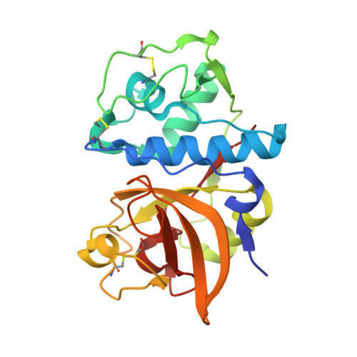
    3KW9, 3KWZ, 3KX1

  • PubMed Abstract: 

    Morphing structural features of HTS-derived chemotypes led to the discovery of novel 2-cyano-pyrimidine inhibitors of cathepsin K with good pharmacokinetic profiles, for example, compound 20 showed high catK potency (IC(50)=4nM), >580-fold selectivity over catL and catB, and oral bioavailability in the rat of 52%.


  • Organizational Affiliation
    • Schering-Plough Corporation, Newhouse, Lanarkshire, ML1 5SH Scotland, United Kingdom. zoran.rankovic@spcorp.com

Macromolecule Content 

  • Total Structure Weight: 24.04 kDa 
  • Atom Count: 1,892 
  • Modeled Residue Count: 213 
  • Deposited Residue Count: 215 
  • Unique protein chains: 1

Macromolecules

Find similar proteins by:|  3D Structure
Entity ID: 1
MoleculeChains  Sequence LengthOrganismDetailsImage
Cathepsin K215Homo sapiensMutation(s): 0 
EC: 3.4.22.38
UniProt & NIH Common Fund Data Resources
Find proteins for P43235 (Homo sapiens)
Explore P43235 
Go to UniProtKB:  P43235
PHAROS:  P43235
GTEx:  ENSG00000143387 
Entity Groups
Sequence Clusters30% Identity50% Identity70% Identity90% Identity95% Identity100% Identity
UniProt GroupP43235
Sequence Annotations
Expand
Reference Sequence

Small Molecules

Binding Affinity Annotations 
IDSourceBinding Affinity
KX1 BindingDB:  3KX1 IC50: 100 (nM) from 1 assay(s)

Experimental Data & Validation

Experimental Data

  • Method: X-RAY DIFFRACTION
  • Resolution: 1.51 Å
  • R-Value Free:  0.306 (Depositor), 0.302 (DCC) 
  • R-Value Work:  0.239 (Depositor), 0.239 (DCC) 
  • R-Value Observed: 0.243 (Depositor) 
Space Group: P 43 21 2
Unit Cell:
Length ( Å )Angle ( ˚ )
a = 56.069α = 90
b = 56.069β = 90
c = 128.829γ = 90
Software Package:
Software NamePurpose
CrystalCleardata collection
REFMACrefinement
CrystalCleardata reduction
CrystalCleardata scaling

Structure Validation

View Full Validation Report


Ligand Structure Quality Assessment 


Entry History 

Deposition Data

Revision History  (Full details and data files)

  • Version 1.0: 2010-03-02
    Type: Initial release
  • Version 1.1: 2011-07-13
    Changes: Version format compliance
  • Version 1.2: 2014-02-12
    Changes: Database references
  • Version 1.3: 2024-04-03
    Changes: Data collection, Database references, Derived calculations, Refinement description
  • Version 1.4: 2024-10-30
    Changes: Structure summary