3KWV

Structural basis for the unfolding of anthrax lethal factor by protective antigen oligomers


Experimental Data Snapshot

  • Method: X-RAY DIFFRACTION
  • Resolution: 3.10 Å
  • R-Value Free: 0.281 
  • R-Value Work: 0.249 
  • R-Value Observed: 0.250 

wwPDB Validation   3D Report Full Report


This is version 1.3 of the entry. See complete history


Literature

Structural basis for the unfolding of anthrax lethal factor by protective antigen oligomers.

Feld, G.K.Thoren, K.L.Kintzer, A.F.Sterling, H.J.Tang, I.I.Greenberg, S.G.Williams, E.R.Krantz, B.A.

(2010) Nat Struct Mol Biol 17: 1383-1390

  • DOI: https://doi.org/10.1038/nsmb.1923
  • Primary Citation of Related Structures:  
    3KWV

  • PubMed Abstract: 

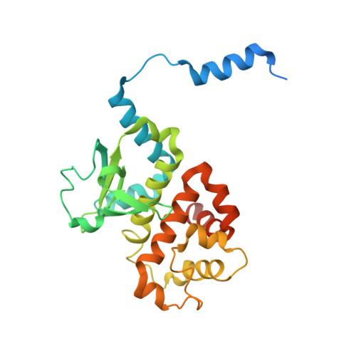
    The protein transporter anthrax lethal toxin is composed of protective antigen (PA), a transmembrane translocase, and lethal factor (LF), a cytotoxic enzyme. After its assembly into holotoxin complexes, PA forms an oligomeric channel that unfolds LF and translocates it into the host cell. We report the crystal structure of the core of a lethal toxin complex to 3.1-Å resolution; the structure contains a PA octamer bound to four LF PA-binding domains (LF(N)). The first α-helix and β-strand of each LF(N) unfold and dock into a deep amphipathic cleft on the surface of the PA octamer, which we call the α clamp. The α clamp possesses nonspecific polypeptide binding activity and is functionally relevant to efficient holotoxin assembly, PA octamer formation, and LF unfolding and translocation. This structure provides insight into the mechanism of translocation-coupled protein unfolding.


  • Organizational Affiliation

    Department of Chemistry, University of California, Berkeley, California, USA.


Macromolecules
Find similar proteins by:  (by identity cutoff)  |  3D Structure
Entity ID: 1
MoleculeChains Sequence LengthOrganismDetailsImage
Protective antigen PA-63
A, B, D, E
548Bacillus anthracisMutation(s): 2 
Gene Names: pagA
Membrane Entity: Yes 
UniProt
Find proteins for P13423 (Bacillus anthracis)
Explore P13423 
Go to UniProtKB:  P13423
Entity Groups  
Sequence Clusters30% Identity50% Identity70% Identity90% Identity95% Identity100% Identity
UniProt GroupP13423
Sequence Annotations
Expand
  • Reference Sequence
Find similar proteins by:  (by identity cutoff)  |  3D Structure
Entity ID: 2
MoleculeChains Sequence LengthOrganismDetailsImage
Lethal factor
C, F
263Bacillus anthracisMutation(s): 0 
Gene Names: lef
EC: 3.4.24.83
Membrane Entity: Yes 
UniProt
Find proteins for P15917 (Bacillus anthracis)
Explore P15917 
Go to UniProtKB:  P15917
Entity Groups  
Sequence Clusters30% Identity50% Identity70% Identity90% Identity95% Identity100% Identity
UniProt GroupP15917
Sequence Annotations
Expand
  • Reference Sequence
Experimental Data & Validation

Experimental Data

  • Method: X-RAY DIFFRACTION
  • Resolution: 3.10 Å
  • R-Value Free: 0.281 
  • R-Value Work: 0.249 
  • R-Value Observed: 0.250 
  • Space Group: P 4 21 2
Unit Cell:
Length ( Å )Angle ( ˚ )
a = 178.381α = 90
b = 178.381β = 90
c = 240.363γ = 90
Software Package:
Software NamePurpose
HKL-2000data collection
PHASERphasing
PHENIXrefinement
HKL-2000data reduction
HKL-2000data scaling

Structure Validation

View Full Validation Report


Entry History 

Deposition Data

Revision History  (Full details and data files)

  • Version 1.0: 2010-11-10
    Type: Initial release
  • Version 1.1: 2011-07-13
    Changes: Version format compliance
  • Version 1.2: 2021-10-13
    Changes: Database references, Derived calculations
  • Version 1.3: 2023-09-06
    Changes: Data collection, Refinement description