3KU3

Crystal structure of a H2N2 influenza virus hemagglutinin, avian like


Experimental Data Snapshot

  • Method: X-RAY DIFFRACTION
  • Resolution: 1.60 Å
  • R-Value Free: 0.230 
  • R-Value Work: 0.199 
  • R-Value Observed: 0.201 

wwPDB Validation   3D Report Full Report


Ligand Structure Quality Assessment 


This is version 2.0 of the entry. See complete history


Literature

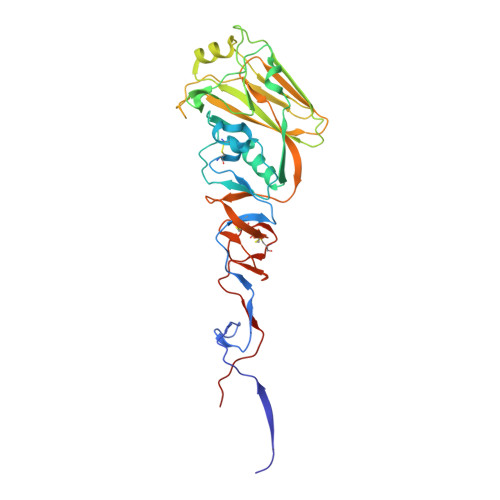
Structure, receptor binding, and antigenicity of influenza virus hemagglutinins from the 1957 H2N2 pandemic.

Xu, R.McBride, R.Paulson, J.C.Basler, C.F.Wilson, I.A.

(2010) J Virol 84: 1715-1721

  • DOI: https://doi.org/10.1128/JVI.02162-09
  • Primary Citation of Related Structures:  
    3KU3

  • PubMed Abstract: 

    The hemagglutinin (HA) envelope protein of influenza viruses mediates essential viral functions, including receptor binding and membrane fusion, and is the major viral antigen for antibody neutralization. The 1957 H2N2 subtype (Asian flu) was one of the three great influenza pandemics of the last century and caused 1 million deaths globally from 1957 to 1968. Three crystal structures of 1957 H2 HAs have been determined at 1.60 to 1.75 A resolutions to investigate the structural basis for their antigenicity and evolution from avian to human binding specificity that contributed to its introduction into the human population. These structures, which represent the highest resolutions yet recorded for a complete ectodomain of a glycosylated viral surface antigen, along with the results of glycan microarray binding analysis, suggest that a hydrophobicity switch at residue 226 and elongation of receptor-binding sites were both critical for avian H2 HA to acquire human receptor specificity. H2 influenza viruses continue to circulate in birds and pigs and, therefore, remain a substantial threat for transmission to humans. The H2 HA structure also reveals a highly conserved epitope that could be harnessed in the design of a broader and more universal influenza A virus vaccine.


  • Organizational Affiliation

    Department of Molecular Biology, BCC-206, The Scripps Research Institute, 10550 North Torrey Pines Road, La Jolla, CA 92037, USA.


Macromolecules
Find similar proteins by:  (by identity cutoff)  |  3D Structure
Entity ID: 1
MoleculeChains Sequence LengthOrganismDetailsImage
Hemagglutinin HA1 chain327Influenza A virus (A/Japan/305/1957(H2N2))Mutation(s): 0 
Gene Names: HAhemagglutinin
UniProt
Find proteins for C7S226 (Influenza A virus (strain A/Japan/305/1957 H2N2))
Explore C7S226 
Go to UniProtKB:  C7S226
Entity Groups  
Sequence Clusters30% Identity50% Identity70% Identity90% Identity95% Identity100% Identity
UniProt GroupC7S226
Sequence Annotations
Expand
  • Reference Sequence
Find similar proteins by:  (by identity cutoff)  |  3D Structure
Entity ID: 2
MoleculeChains Sequence LengthOrganismDetailsImage
Hemagglutinin HA2 chain174Influenza A virus (A/Japan/305/1957(H2N2))Mutation(s): 0 
Gene Names: HAhemagglutinin
UniProt
Find proteins for C7S226 (Influenza A virus (strain A/Japan/305/1957 H2N2))
Explore C7S226 
Go to UniProtKB:  C7S226
Entity Groups  
Sequence Clusters30% Identity50% Identity70% Identity90% Identity95% Identity100% Identity
UniProt GroupC7S226
Sequence Annotations
Expand
  • Reference Sequence
Oligosaccharides

Help

Entity ID: 3
MoleculeChains Length2D Diagram Glycosylation3D Interactions
2-acetamido-2-deoxy-beta-D-glucopyranose-(1-4)-2-acetamido-2-deoxy-beta-D-glucopyranose
C, D
2N-Glycosylation
Glycosylation Resources
GlyTouCan:  G42666HT
GlyCosmos:  G42666HT
GlyGen:  G42666HT
Small Molecules
Experimental Data & Validation

Experimental Data

  • Method: X-RAY DIFFRACTION
  • Resolution: 1.60 Å
  • R-Value Free: 0.230 
  • R-Value Work: 0.199 
  • R-Value Observed: 0.201 
  • Space Group: P 63
Unit Cell:
Length ( Å )Angle ( ˚ )
a = 70.252α = 90
b = 70.252β = 90
c = 236.879γ = 120
Software Package:
Software NamePurpose
DENZOdata reduction
SCALEPACKdata scaling
PHASERphasing
REFMACrefinement
PDB_EXTRACTdata extraction
Blu-Icedata collection
HKL-2000data reduction
HKL-2000data scaling

Structure Validation

View Full Validation Report


Ligand Structure Quality Assessment 


Entry History 

Deposition Data

Revision History  (Full details and data files)

  • Version 1.0: 2010-01-19
    Type: Initial release
  • Version 1.1: 2011-07-13
    Changes: Advisory, Refinement description, Version format compliance
  • Version 1.2: 2017-11-01
    Changes: Refinement description
  • Version 2.0: 2020-07-29
    Type: Remediation
    Reason: Carbohydrate remediation
    Changes: Atomic model, Data collection, Database references, Derived calculations, Structure summary