3KTM | pdb_00003ktm

Structure of the Heparin-induced E1-Dimer of the Amyloid Precursor Protein (APP)


Experimental Data Snapshot

  • Method: X-RAY DIFFRACTION
  • Resolution: 2.70 Å
  • R-Value Free: 
    0.250 (Depositor), 0.249 (DCC) 
  • R-Value Work: 
    0.215 (Depositor), 0.215 (DCC) 

Starting Models: experimental
View more details

wwPDB Validation 3D Report Full Report

Validation slider image for 3KTM

This is version 1.3 of the entry. See complete history

Literature

Structure and biochemical analysis of the heparin-induced E1 dimer of the amyloid precursor protein.

Dahms, S.O.Hoefgen, S.Roeser, D.Schlott, B.Guhrs, K.H.Than, M.E.

(2010) Proc Natl Acad Sci U S A 107: 5381-5386

  • DOI: https://doi.org/10.1073/pnas.0911326107
  • Primary Citation Related Structures: 
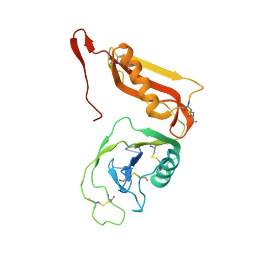
    3KTM

  • PubMed Abstract: 

    The amyloid precursor protein (APP) is the key player in Alzheimer's disease pathology, yet APP and its analogues are also essential for neuronal development and cell homeostasis in mammals. We have determined the crystal structure of the entire N-terminal APP-E1 domain consisting of the growth factor like and the copper binding domains at 2.7-A resolution and show that E1 functions as a rigid functional entity. The two subdomains interact tightly in a pH-dependent manner via an evolutionarily conserved interface area. Two E1 entities dimerize upon their interaction with heparin, requiring 8-12 sugar rings to form the heparin-bridged APP-E1 dimer in an endothermic and pH-dependent process that is characterized by a low micromolar dissociation constant. Limited proteolysis confirms that the heparin-bridged E1 dimers obtained in solution correspond to a dimer contact in our crystal, enabling us to model this heparin-[APP-E1](2) complex. Correspondingly, the APP-based signal transduction, cell-cell- and/or cell-ECM interaction should depend on dimerization induced by heparin, as well as on pH, arguing that APP could fulfill different functions depending on its (sub)cellular localization.


  • Organizational Affiliation
    • Leibniz Institute for Age Research-Fritz Lipman Institute, Protein Crystallography Group, Jena, Germany.

Macromolecule Content 

  • Total Structure Weight: 175.53 kDa 
  • Atom Count: 11,436 
  • Modeled Residue Count: 1,396 
  • Deposited Residue Count: 1,528 
  • Unique protein chains: 1

Macromolecules

Find similar proteins by:|  3D Structure
Entity ID: 1
MoleculeChains  Sequence LengthOrganismDetailsImage
Amyloid beta A4 protein
A, B, C, D, E
A, B, C, D, E, F, G, H
191Homo sapiensMutation(s): 0 
Gene Names: APPA4AD1
UniProt & NIH Common Fund Data Resources
Find proteins for P05067 (Homo sapiens)
Explore P05067 
Go to UniProtKB:  P05067
PHAROS:  P05067
GTEx:  ENSG00000142192 
Entity Groups
Sequence Clusters30% Identity50% Identity70% Identity90% Identity95% Identity100% Identity
UniProt GroupP05067
Sequence Annotations
Expand
Reference Sequence

Small Molecules

Ligands 3 Unique
IDChains Name / Formula / InChI Key2D Diagram3D Interactions
SO4

Query on SO4


Download:Ideal Coordinates CCD File
K [auth B],
N [auth C],
Q [auth D],
S [auth E],
X [auth H]
SULFATE ION
O4 S
QAOWNCQODCNURD-UHFFFAOYSA-L
BU4

Query on BU4


Download:Ideal Coordinates CCD File
I [auth A]
J [auth B]
M [auth C]
P [auth D]
R [auth E]
I [auth A],
J [auth B],
M [auth C],
P [auth D],
R [auth E],
U [auth F],
V [auth G],
W [auth H]
(3R)-butane-1,3-diol
C4 H10 O2
PUPZLCDOIYMWBV-SCSAIBSYSA-N
ACT

Query on ACT


Download:Ideal Coordinates CCD File
L [auth B],
O [auth C],
T [auth E],
Y [auth H]
ACETATE ION
C2 H3 O2
QTBSBXVTEAMEQO-UHFFFAOYSA-M

Experimental Data & Validation

Experimental Data

  • Method: X-RAY DIFFRACTION
  • Resolution: 2.70 Å
  • R-Value Free:  0.250 (Depositor), 0.249 (DCC) 
  • R-Value Work:  0.215 (Depositor), 0.215 (DCC) 
Space Group: H 3
Unit Cell:
Length ( Å )Angle ( ˚ )
a = 143.99α = 90
b = 143.99β = 90
c = 351.19γ = 120
Software Package:
Software NamePurpose
MxCuBEdata collection
PHASERphasing
CNSrefinement
MOSFLMdata reduction
SCALAdata scaling

Structure Validation

View Full Validation Report


Entry History 

Deposition Data

Revision History  (Full details and data files)

  • Version 1.0: 2010-02-23
    Type: Initial release
  • Version 1.1: 2011-07-13
    Changes: Version format compliance
  • Version 1.2: 2023-09-06
    Changes: Data collection, Database references, Derived calculations, Refinement description
  • Version 1.3: 2024-10-30
    Changes: Structure summary