3KSG | pdb_00003ksg

structure of fRMsr of Staphylococcus aureus (complex with substrate)


Experimental Data Snapshot

  • Method: X-RAY DIFFRACTION
  • Resolution: 2.30 Å
  • R-Value Free: 
    0.252 (Depositor), 0.247 (DCC) 
  • R-Value Work: 
    0.222 (Depositor), 0.221 (DCC) 
  • R-Value Observed: 
    0.222 (Depositor) 

Starting Model: experimental
View more details

wwPDB Validation 3D Report Full Report

Validation slider image for 3KSG

Ligand Structure Quality Assessment 


This is version 1.2 of the entry. See complete history

Literature

Structure of fRMsr of Staphylococcus aureus

Bong, S.M.Chi, Y.M.

To be published.

Macromolecule Content 

  • Total Structure Weight: 36.19 kDa 
  • Atom Count: 2,455 
  • Modeled Residue Count: 306 
  • Deposited Residue Count: 320 
  • Unique protein chains: 1

Macromolecules

Find similar proteins by:|  3D Structure
Entity ID: 1
MoleculeChains  Sequence LengthOrganismDetailsImage
Putative uncharacterized protein
A, B
160Staphylococcus aureus subsp. aureus MRSA252Mutation(s): 1 
Gene Names: SAR1796
EC: 1.8.4.14
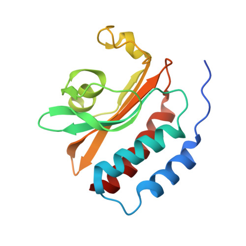
Experimental Data & Validation

Experimental Data

  • Method: X-RAY DIFFRACTION
  • Resolution: 2.30 Å
  • R-Value Free:  0.252 (Depositor), 0.247 (DCC) 
  • R-Value Work:  0.222 (Depositor), 0.221 (DCC) 
  • R-Value Observed: 0.222 (Depositor) 
Space Group: P 1 21 1
Unit Cell:
Length ( Å )Angle ( ˚ )
a = 41.582α = 90
b = 87.492β = 101.25
c = 42.862γ = 90
Software Package:
Software NamePurpose
ADSCdata collection
MOLREPphasing
CNSrefinement
HKL-2000data reduction
HKL-2000data scaling

Structure Validation

View Full Validation Report


Ligand Structure Quality Assessment 


Entry History 

Deposition Data

Revision History  (Full details and data files)

  • Version 1.0: 2010-05-26
    Type: Initial release
  • Version 1.1: 2011-07-13
    Changes: Version format compliance
  • Version 1.2: 2023-11-01
    Changes: Data collection, Database references, Derived calculations, Refinement description