3KSC | pdb_00003ksc

Crystal structure of pea prolegumin, an 11S seed globulin from Pisum sativum L.


Experimental Data Snapshot

  • Method: X-RAY DIFFRACTION
  • Resolution: 2.61 Å
  • R-Value Free: 
    0.234 (Depositor), 0.229 (DCC) 
  • R-Value Work: 
    0.175 (Depositor), 0.172 (DCC) 
  • R-Value Observed: 
    0.178 (Depositor) 

Starting Model: experimental
View more details

wwPDB Validation 3D Report Full Report

Validation slider image for 3KSC

This is version 1.4 of the entry. See complete history

Literature

Conservation and divergence on plant seed 11S globulins based on crystal structures.

Tandang-Silvas, M.R.Fukuda, T.Fukuda, C.Prak, K.Cabanos, C.Kimura, A.Itoh, T.Mikami, B.Utsumi, S.Maruyama, N.

(2010) Biochim Biophys Acta 1804: 1432-1442

  • DOI: https://doi.org/10.1016/j.bbapap.2010.02.016
  • Primary Citation Related Structures: 
    2D5F, 2D5H, 2E9Q, 3KGL, 3KSC

  • PubMed Abstract: 

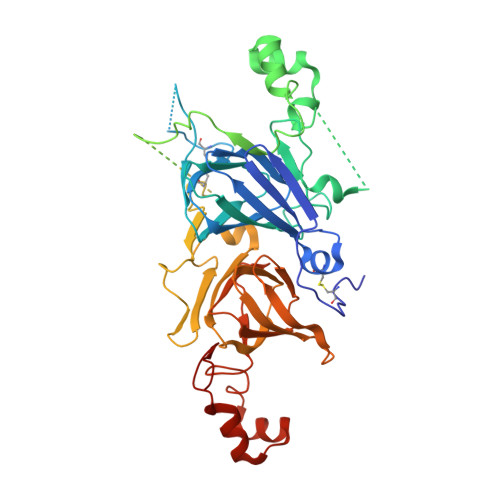
    The crystal structures of two pro-11S globulins namely: rapeseed procruciferin and pea prolegumin are presented here. We have extensively compared them with the other known structures of plant seed 11S and 7S globulins. In general, the disordered regions in the crystal structures among the 11S globulins correspond to their five variable regions. Variable region III of procruciferin is relatively short and is in a loop conformation. This region is highly disordered in other pro-11S globulin crystals. Local helical and strand variations also occur across the group despite general structure conservation. We showed how these variations may alter specific physicochemical, functional and physiological properties. Aliphatic hydrophobic residues on the molecular surface correlate well with Tm values of the globulins. We also considered other structural features that were reported to influence thermal stability but no definite conclusion was drawn since each factor has additive or subtractive effect. Comparison between proA3B4 and mature A3B4 revealed an increase in r.m.s.d. values near variable regions II and IV. Both regions are on the IE face. Secondary structure based alignment of 11S and 7S globulins revealed 16 identical residues. Based on proA3B4 sequence, Pro60, Gly128, Phe163, Phe208, Leu213, Leu227, Ile237, Pro382, Val404, Pro425 and Val 466 are involved in trimer formation and stabilization. Gly28, Gly74, Asp135, Gly349 and Gly397 are involved in correct globular folding.


  • Organizational Affiliation
    • Laboratory of Food Quality Design and Development, Graduate School of Agriculture, Kyoto University, Uji, Kyoto, Kyoto 611-0011, Japan.

Macromolecule Content 

  • Total Structure Weight: 342.92 kDa 
  • Atom Count: 18,563 
  • Modeled Residue Count: 2,274 
  • Deposited Residue Count: 2,976 
  • Unique protein chains: 1

Macromolecules

Find similar proteins by:|  3D Structure
Entity ID: 1
MoleculeChains  Sequence LengthOrganismDetailsImage
LegA class
A, B, C, D, E
A, B, C, D, E, F
496Lathyrus oleraceusMutation(s): 0 
Gene Names: legAPlant gene
UniProt
Find proteins for P02857 (Pisum sativum)
Explore P02857 
Go to UniProtKB:  P02857
Entity Groups
Sequence Clusters30% Identity50% Identity70% Identity90% Identity95% Identity100% Identity
UniProt GroupP02857
Sequence Annotations
Expand
Reference Sequence

Small Molecules

Ligands 2 Unique
IDChains Name / Formula / InChI Key2D Diagram3D Interactions
SO4

Query on SO4


Download:Ideal Coordinates CCD File
BA [auth D]
FA [auth E]
G [auth A]
GA [auth E]
H [auth A]
BA [auth D],
FA [auth E],
G [auth A],
GA [auth E],
H [auth A],
K [auth B],
L [auth B],
M [auth B],
N [auth B],
O [auth B],
P [auth B],
V [auth C],
W [auth C]
SULFATE ION
O4 S
QAOWNCQODCNURD-UHFFFAOYSA-L
GOL

Query on GOL


Download:Ideal Coordinates CCD File
AA [auth C]
CA [auth D]
DA [auth D]
EA [auth D]
HA [auth E]
AA [auth C],
CA [auth D],
DA [auth D],
EA [auth D],
HA [auth E],
I [auth A],
IA [auth F],
J [auth A],
JA [auth F],
KA [auth F],
Q [auth B],
R [auth B],
S [auth B],
T [auth B],
U [auth B],
X [auth C],
Y [auth C],
Z [auth C]
GLYCEROL
C3 H8 O3
PEDCQBHIVMGVHV-UHFFFAOYSA-N

Experimental Data & Validation

Experimental Data

  • Method: X-RAY DIFFRACTION
  • Resolution: 2.61 Å
  • R-Value Free:  0.234 (Depositor), 0.229 (DCC) 
  • R-Value Work:  0.175 (Depositor), 0.172 (DCC) 
  • R-Value Observed: 0.178 (Depositor) 
Space Group: P 21 21 21
Unit Cell:
Length ( Å )Angle ( ˚ )
a = 129.777α = 90
b = 148.441β = 90
c = 149.894γ = 90
Software Package:
Software NamePurpose
PHENIXrefinement
HKL-2000data reduction
HKL-2000data scaling

Structure Validation

View Full Validation Report


Entry History 

Deposition Data

Revision History  (Full details and data files)

  • Version 1.0: 2010-04-21
    Type: Initial release
  • Version 1.1: 2011-07-13
    Changes: Version format compliance
  • Version 1.2: 2013-12-11
    Changes: Database references
  • Version 1.3: 2023-11-01
    Changes: Data collection, Database references, Derived calculations, Refinement description
  • Version 1.4: 2024-11-06
    Changes: Structure summary