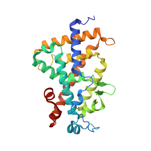
3KPZ | pdb_00003kpz

Crystal structure of a novel vitamin D3 analogue, ZK203278 showing dissociated profile


Experimental Data Snapshot

  • Method: X-RAY DIFFRACTION
  • Resolution: 1.90 Å
  • R-Value Free: 
    0.228 (Depositor), 0.230 (DCC) 
  • R-Value Work: 
    0.195 (Depositor), 0.200 (DCC) 
  • R-Value Observed: 
    0.228 (Depositor) 

Starting Model: experimental
View more details

wwPDB Validation   3D Report Full Report


Ligand Structure Quality Assessment 

Created with Raphaël 2.3.0Worse 01 BetterLigand structure goodness of fit to experimental dataBest fitted ZNEClick on this verticalbar to view details

This is version 1.3 of the entry. See complete history


Literature

Crystal structure of a vitamin D3 analog, ZK203278, showing dissociated profile.

Rochel, N.Moras, D.

(2012) Anticancer Res 32: 335-339

  • Primary Citation of Related Structures:  
    3KPZ

  • PubMed Abstract: 

    The plethora of actions of 1α,25-dihydroxyvitamin D(3), the active form of the seco-steroid hormone vitamin D, in various systems suggested wide clinical applications in treatments for renal osteodystrophy, osteoporosis, psoriasis, cancer, autoimmune diseases and prevention of graft rejection. However, the major side-effects of hypercalcemia of VDR ligands limit their use. ZK203278, a novel synthetic analog has been shown to act as a potent immunomodulator and presents dissociated biologic profile with low calcemic side-effects. Here, we described the crystal structures of the hVDR ligand-binding domain in complex with ZK203278 and determined its correlation with its specific dissociated biologic profile. The VDR/ZK203278 structure, in comparison with VDR/1α,25-dihydroxyvitamin D(3), shows specific interactions of the thiazole group of ZK203278 with residues of H3, H11 and H12. These specific interactions may lead to altered selective interactions with co-regulators and consequently to the dissociated biologic profile of this novel ligand.


  • Organizational Affiliation

    Institut de Génétique et de Biologie Moléculaire et Cellulaire (IGBMC), Institut National de Santé et de Recherche Médicale (INSERM) U964/Centre National de Recherche Scientifique (CNRS) UMR 7104/Université de Strasbourg, 67404 Illkirch, France. rochel@igbmc.fr


Macromolecules
Find similar proteins by:  (by identity cutoff)  |  3D Structure
Entity ID: 1
MoleculeChains Sequence LengthOrganismDetailsImage
Vitamin D3 receptor259Homo sapiensMutation(s): 0 
Gene Names: VDRNR1I1
UniProt & NIH Common Fund Data Resources
Find proteins for P11473 (Homo sapiens)
Explore P11473 
Go to UniProtKB:  P11473
PHAROS:  P11473
GTEx:  ENSG00000111424 
Entity Groups  
Sequence Clusters30% Identity50% Identity70% Identity90% Identity95% Identity100% Identity
UniProt GroupP11473
Sequence Annotations
Expand
  • Reference Sequence
Small Molecules
Ligands 1 Unique
IDChains Name / Formula / InChI Key2D Diagram3D Interactions
ZNE
Query on ZNE

Download Ideal Coordinates CCD File 
B [auth A](1R,3S,5Z)-5-[(2E)-2-{(1R,3aS,7aR)-1-[(1R,5S)-5-hydroxy-1-methyl-5-(1,3-thiazol-2-yl)pentyl]-7a-methyloctahydro-4H-inden-4-ylidene}ethylidene]-4-methylidenecyclohexane-1,3-diol
C28 H41 N O3 S
AZVJHDPZXYTKQV-LCRUICLWSA-N
Experimental Data & Validation

Experimental Data

  • Method: X-RAY DIFFRACTION
  • Resolution: 1.90 Å
  • R-Value Free:  0.228 (Depositor), 0.230 (DCC) 
  • R-Value Work:  0.195 (Depositor), 0.200 (DCC) 
  • R-Value Observed: 0.228 (Depositor) 
Space Group: P 21 21 21
Unit Cell:
Length ( Å )Angle ( ˚ )
a = 44.887α = 90
b = 51.892β = 90
c = 132.204γ = 90
Software Package:
Software NamePurpose
CNSrefinement
PDB_EXTRACTdata extraction
HKL-2000data reduction
HKL-2000data scaling
CNSphasing

Structure Validation

View Full Validation Report


Ligand Structure Quality Assessment 

Created with Raphaël 2.3.0Worse 01 BetterLigand structure goodness of fit to experimental dataBest fitted ZNEClick on this verticalbar to view details

Entry History 

Deposition Data

Revision History  (Full details and data files)

  • Version 1.0: 2011-10-12
    Type: Initial release
  • Version 1.1: 2011-10-26
    Changes: Structure summary
  • Version 1.2: 2012-04-25
    Changes: Database references
  • Version 1.3: 2023-09-06
    Changes: Data collection, Database references, Derived calculations, Refinement description