Optimization of piperidyl-ureas as inhibitors of soluble epoxide hydrolase.
Eldrup, A.B., Soleymanzadeh, F., Farrow, N.A., Kukulka, A., De Lombaert, S.(2010) Bioorg Med Chem Lett 20: 571-575
- PubMed: 19969453 Search on PubMed
- DOI: https://doi.org/10.1016/j.bmcl.2009.11.091
- Primary Citation Related Structures:
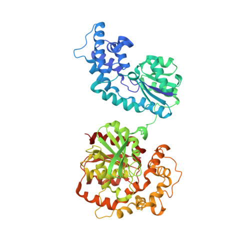
3KOO - PubMed Abstract:
Inhibition of sEH is hypothesized to lead to an increase in epoxyeicosatrienoic acids resulting in the potentiation of their anti-inflammatory and vasodilatory effects. In an effort to explore sEH inhibition as an avenue for the development of vasodilatory and cardio- or renal-protective agents, a lead identified through high-throughput screening was optimized, guided by the determination of a solid state co-structure with sEH. Replacement of potential toxicophores was followed by optimization of cell-based potency and ADME properties to provide a new class of functionally potent sEH inhibitors with attractive in vitro metabolic profiles and high and sustained plasma exposures after oral administration in the rat.
- Department of Medicinal Chemistry, Boehringer Ingelheim Pharmaceuticals, Ridgefield, CT 06877, United States. anne.eldrup@boehringer-ingelheim.com
Organizational Affiliation: