3KO8

Crystal Structure of UDP-galactose 4-epimerase


Experimental Data Snapshot

  • Method: X-RAY DIFFRACTION
  • Resolution: 1.80 Å
  • R-Value Free: 0.225 
  • R-Value Work: 0.204 
  • R-Value Observed: 0.205 

wwPDB Validation   3D Report Full Report


Ligand Structure Quality Assessment 


This is version 1.3 of the entry. See complete history


Literature

Crystal structure of UDP-galactose 4-epimerase from the hyperthermophilic archaeon Pyrobaculum calidifontis

Sakuraba, H.Kawai, T.Yoneda, K.Ohshima, T.

(2011) Arch Biochem Biophys 512: 126-134

  • DOI: https://doi.org/10.1016/j.abb.2011.05.013
  • Primary Citation of Related Structures:  
    3KO8

  • PubMed Abstract: 

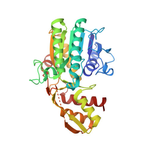
    The crystal structure of a highly thermostable UDP-galactose 4-epimerase (GalE) from the hyperthermophilic archaeon Pyrobaculum calidifontis was determined at a resolution of 1.8Å. The asymmetric unit contained one subunit, and the functional dimer was generated by a crystallographic two-fold axis. Each monomer consisted of a Rossmann-fold domain with NAD bound and a carboxyl terminal domain. The overall structure of P. calidifontis GalE showed significant similarity to the structures of the GalEs from Escherichia coli, human and Trypanosoma brucei. However, the sizes of several surface loops were markedly smaller in P. calidifontis GalE than the corresponding loops in the other enzymes. Structural comparison revealed that the presence of an extensive hydrophobic interaction at the subunit interface is likely the main factor contributing to the hyperthermostability of the P. calidifontis enzyme. Within the NAD-binding site of P. calidifontis GalE, a loop (NAD-binding loop) tightly holds the adenine ribose moiety of NAD. Moreover, a deletion mutant lacking this loop bound NAD in a loose, reversible manner. Thus the presence of the NAD-binding loop in GalE is largely responsible for preventing the release of the cofactor from the holoenzyme.


  • Organizational Affiliation

    Department of Applied Biological Science, Kagawa University, Ikenobe, Miki-cho, Kita-gun, Japan.


Macromolecules
Find similar proteins by:  (by identity cutoff)  |  3D Structure
Entity ID: 1
MoleculeChains Sequence LengthOrganismDetailsImage
NAD-dependent epimerase/dehydratase312Pyrobaculum calidifontis JCM 11548Mutation(s): 0 
EC: 5.1.3.2
UniProt
Find proteins for A3MUJ4 (Pyrobaculum calidifontis (strain DSM 21063 / JCM 11548 / VA1))
Explore A3MUJ4 
Go to UniProtKB:  A3MUJ4
Entity Groups  
Sequence Clusters30% Identity50% Identity70% Identity90% Identity95% Identity100% Identity
UniProt GroupA3MUJ4
Sequence Annotations
Expand
  • Reference Sequence
Small Molecules
Ligands 2 Unique
IDChains Name / Formula / InChI Key2D Diagram3D Interactions
NAD
Query on NAD

Download Ideal Coordinates CCD File 
B [auth A]NICOTINAMIDE-ADENINE-DINUCLEOTIDE
C21 H27 N7 O14 P2
BAWFJGJZGIEFAR-NNYOXOHSSA-N
PO4
Query on PO4

Download Ideal Coordinates CCD File 
C [auth A]PHOSPHATE ION
O4 P
NBIIXXVUZAFLBC-UHFFFAOYSA-K
Experimental Data & Validation

Experimental Data

  • Method: X-RAY DIFFRACTION
  • Resolution: 1.80 Å
  • R-Value Free: 0.225 
  • R-Value Work: 0.204 
  • R-Value Observed: 0.205 
  • Space Group: P 41 21 2
Unit Cell:
Length ( Å )Angle ( ˚ )
a = 100.803α = 90
b = 100.803β = 90
c = 76.302γ = 90
Software Package:
Software NamePurpose
MOLREPphasing
CNSrefinement
PDB_EXTRACTdata extraction
HKL-2000data collection
HKL-2000data reduction
HKL-2000data scaling

Structure Validation

View Full Validation Report


Ligand Structure Quality Assessment 


Entry History 

Deposition Data

Revision History  (Full details and data files)

  • Version 1.0: 2010-11-17
    Type: Initial release
  • Version 1.1: 2011-07-13
    Changes: Version format compliance
  • Version 1.2: 2011-08-03
    Changes: Database references
  • Version 1.3: 2023-11-01
    Changes: Data collection, Database references, Derived calculations, Refinement description