Crystal Structure of the ATP-Dependent Maturation Factor of Ni,Fe-Containing Carbon Monoxide Dehydrogenases
Jeoung, J.H., Giese, T., Grunwald, M., Dobbek, H.(2010) J Mol Biology 396: 1165-1179
- PubMed: 20064527 Search on PubMed
- DOI: https://doi.org/10.1016/j.jmb.2009.12.062
- Primary Citation Related Structures:
3KJE, 3KJG, 3KJH, 3KJI - PubMed Abstract:
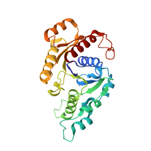
CooC proteins are ATPases involved in the incorporation of nickel into the complex active site ([Ni-4Fe-4S]) cluster of Ni,Fe-dependent carbon monoxide dehydrogenases. The genome of the carboxydotrophic bacterium Carboxydothermus hydrogenoformans encodes five carbon monoxide dehydrogenases and three CooC-type proteins, of which CooC1 was shown to be a nickel-binding ATPase. We determined the crystal structure of CooC1 in four different states: empty, ADP-bound, Zn(2+)/ADP-bound, and Zn(2+)-bound. The structure of CooC1 consists of two spatially separated functional modules: an ATPase module containing the deviant Walker A motif and a metal-binding module that confers the specific function of CooC1. The ATPase module is homologous to other members of the MinD family and, in analogy to the dimeric structure of ATP-bound Soj, is likely responsible for the ATP-dependent dimerization of CooC1. Its core topology classifies CooC1 as a member of the MinD family of SIMIBI (signal recognition particle, MinD and BioD)-class NTPases. The crystal structure of Zn(2+)-bound CooC1 reveals a conserved C-X-C motif as the metal-binding site responsible for metal-induced dimerization. The competitive binding of Ni(2+) and Zn(2+) to CooC1 in solution confirms that the conserved C-X-C motif is also responsible for the interaction with Ni(2+). A comparison of the different CooC1 structures determined suggests a mutual dependence of metal-binding site and nucleotide-binding site.
- AG Bioanorganische Chemie, Universität Bayreuth, Universitätsstrasse 30, 95447 Bayreuth, Germany.
Organizational Affiliation: