Continued exploration of the triazolopyridine scaffold as a platform for p38 MAP kinase inhibition.
Jerome, K.D., Rucker, P.V., Xing, L., Shieh, H.S., Baldus, J.E., Selness, S.R., Letavic, M.A., Braganza, J.F., McClure, K.F.(2010) Bioorg Med Chem Lett 20: 469-473
- PubMed: 19969459 Search on PubMed
- DOI: https://doi.org/10.1016/j.bmcl.2009.11.114
- Primary Citation Related Structures:
3KF7 - PubMed Abstract:
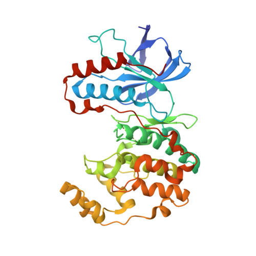
The structure based drug design, synthesis and structure-activity relationship of a series of C6 sulfur linked triazolopyridine based p38 inhibitors are described. The metabolic deficiencies of this series were overcome through changes in the C6 linker from sulfur to methylene, which was predicted by molecular modeling to be bioisosteric. X-ray of the ethylene linked compound 61 confirmed the predicted binding orientation of the scaffold in the p38 enzyme.
- Pfizer Global Research and Development, St. Louis Laboratories, 700 Chesterfield Pkwy. W., Chesterfield, MO 63017, USA. kevin.d.jerome@pfizer.com
Organizational Affiliation: