


Crystal structure of PL kinase in complex with MgATP and PLP: Structural basis of severe induced MgATP substrate inhibition of the enzyme
Gandhi, A.K., Musayev, F.N., Safo, M.K.To be published.
Experimental Data Snapshot
Entity ID: 1 | |||||
|---|---|---|---|---|---|
| Molecule | Chains | Sequence Length | Organism | Details | Image |
| Pyridoxal kinase | 312 | Homo sapiens | Mutation(s): 0 Gene Names: C21orf124, C21orf97, PDXK, PKH, PNK EC: 2.7.1.35 | 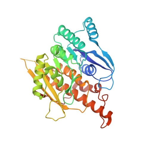 | |
UniProt & NIH Common Fund Data Resources | |||||
PHAROS: O00764 GTEx: ENSG00000160209 | |||||
Entity Groups | |||||
| Sequence Clusters | 30% Identity50% Identity70% Identity90% Identity95% Identity100% Identity | ||||
| UniProt Group | O00764 | ||||
Sequence AnnotationsExpand | |||||
Reference Sequence | |||||
| Ligands 6 Unique | |||||
|---|---|---|---|---|---|
| ID | Chains | Name / Formula / InChI Key | 2D Diagram | 3D Interactions | |
| ATP Download:Ideal Coordinates CCD File | F [auth A], Q [auth B] | ADENOSINE-5'-TRIPHOSPHATE C10 H16 N5 O13 P3 ZKHQWZAMYRWXGA-KQYNXXCUSA-N |  | ||
| PLP Download:Ideal Coordinates CCD File | G [auth A], S [auth B] | PYRIDOXAL-5'-PHOSPHATE C8 H10 N O6 P NGVDGCNFYWLIFO-UHFFFAOYSA-N |  | ||
| MPD Download:Ideal Coordinates CCD File | H [auth A] I [auth A] J [auth A] R [auth B] T [auth B] | (4S)-2-METHYL-2,4-PENTANEDIOL C6 H14 O2 SVTBMSDMJJWYQN-YFKPBYRVSA-N |  | ||
| SO4 Download:Ideal Coordinates CCD File | AA [auth B] BA [auth B] CA [auth B] K [auth A] L [auth A] | SULFATE ION O4 S QAOWNCQODCNURD-UHFFFAOYSA-L |  | ||
| MG Download:Ideal Coordinates CCD File | C [auth A], D [auth A], N [auth B], O [auth B] | MAGNESIUM ION Mg JLVVSXFLKOJNIY-UHFFFAOYSA-N |  | ||
| NA Download:Ideal Coordinates CCD File | E [auth A], P [auth B] | SODIUM ION Na FKNQFGJONOIPTF-UHFFFAOYSA-N |  | ||
| Length ( Å ) | Angle ( ˚ ) |
|---|---|
| a = 91.202 | α = 90 |
| b = 114.585 | β = 90 |
| c = 169.414 | γ = 90 |
| Software Name | Purpose |
|---|---|
| d*TREK | data scaling |
| CNS | refinement |
| PDB_EXTRACT | data extraction |
| CrystalClear | data collection |
| d*TREK | data reduction |


