3KCX | pdb_00003kcx

Factor inhibiting HIF-1 alpha in complex with Clioquinol


Experimental Data Snapshot

  • Method: X-RAY DIFFRACTION
  • Resolution: 2.60 Å
  • R-Value Free: 
    0.292 (Depositor), 0.278 (DCC) 
  • R-Value Work: 
    0.244 (Depositor), 0.236 (DCC) 
  • R-Value Observed: 
    0.246 (Depositor) 

wwPDB Validation 3D Report Full Report

Validation slider image for 3KCX

Ligand Structure Quality Assessment 


This is version 1.4 of the entry. See complete history

Literature

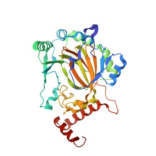
Crystal structures of human FIH-1 in complex with quinol family inhibitors

Moon, H.Han, S.Park, H.Choe, J.

(2010) Mol Cells 29: 471-474

  • DOI: https://doi.org/10.1007/s10059-010-0058-3
  • Primary Citation Related Structures: 
    3KCX, 3KCY

  • PubMed Abstract: 

    Hypoxia-Inducible Factor-1 (HIF-1) plays an important role as a transcription factor under hypoxia. It activates numerous genes including those involved in angiogenesis, glucose metabolisms, cell proliferation and cell survival. The HIF-1 alpha subunit is regulated by 2-oxoglutarate (OG)- and Fe(II)-dependent hydroxylases, including Factor Inhibiting HIF-1 (FIH-1). FIH-1 hydroxylates Asn803 of HIF-1 alpha and blocks its interaction with co-activating molecules. Quinol family compounds such as 5-chloro-7-iodo-8-hydroxyquinoline (Clioquinol) have been shown to inhibit the hydroxylation activity of FIH-1. Here we determined the complex crystal structures of FIH-1: Clioquinol and FIH-1: 8-Hydroxyquinoline. Clioquinol and 8-Hydroxyquinoline bind to the active site of FIH-1 by coordinating the Fe(II) ion, thereby inhibiting the binding of a co-substrate, 2OG. Contrary to other known FIH-1 inhibitors that have negative charges, Clioquinol and 8-hydroxyquinoline are neutral in charge and can provide a template for improved inhibitor design that can selectively inhibit FIH-1.


  • Organizational Affiliation
    • Department of Life Science, University of Seoul, Seoul 130-743, Korea.

Macromolecule Content 

  • Total Structure Weight: 39.98 kDa 
  • Atom Count: 2,806 
  • Modeled Residue Count: 332 
  • Deposited Residue Count: 335 
  • Unique protein chains: 1

Macromolecules

Find similar proteins by:|  3D Structure
Entity ID: 1
MoleculeChains  Sequence LengthOrganismDetailsImage
Hypoxia-inducible factor 1-alpha inhibitor335Homo sapiensMutation(s): 0 
EC: 1.14.11.16 (PDB Primary Data), 1.14.11.30 (UniProt)
UniProt & NIH Common Fund Data Resources
Find proteins for Q9NWT6 (Homo sapiens)
Explore Q9NWT6 
Go to UniProtKB:  Q9NWT6
PHAROS:  Q9NWT6
GTEx:  ENSG00000166135 
Entity Groups
Sequence Clusters30% Identity50% Identity70% Identity90% Identity95% Identity100% Identity
UniProt GroupQ9NWT6
Sequence Annotations
Expand
Reference Sequence

Small Molecules

Ligands 4 Unique
IDChains Name / Formula / InChI Key2D Diagram3D Interactions
CQL

Query on CQL


Download:Ideal Coordinates CCD File
H [auth A]5-chloro-7-iodoquinolin-8-ol
C9 H5 Cl I N O
QCDFBFJGMNKBDO-UHFFFAOYSA-N
SO4

Query on SO4


Download:Ideal Coordinates CCD File
C [auth A],
D [auth A]
SULFATE ION
O4 S
QAOWNCQODCNURD-UHFFFAOYSA-L
GOL

Query on GOL


Download:Ideal Coordinates CCD File
E [auth A],
F [auth A],
G [auth A]
GLYCEROL
C3 H8 O3
PEDCQBHIVMGVHV-UHFFFAOYSA-N
FE2

Query on FE2


Download:Ideal Coordinates CCD File
B [auth A]FE (II) ION
Fe
CWYNVVGOOAEACU-UHFFFAOYSA-N

Experimental Data & Validation

Experimental Data

  • Method: X-RAY DIFFRACTION
  • Resolution: 2.60 Å
  • R-Value Free:  0.292 (Depositor), 0.278 (DCC) 
  • R-Value Work:  0.244 (Depositor), 0.236 (DCC) 
  • R-Value Observed: 0.246 (Depositor) 
Space Group: P 41 21 2
Unit Cell:
Length ( Å )Angle ( ˚ )
a = 86.746α = 90
b = 86.746β = 90
c = 146.525γ = 90
Software Package:
Software NamePurpose
DENZOdata reduction
SCALEPACKdata scaling
REFMACrefinement
PDB_EXTRACTdata extraction

Structure Validation

View Full Validation Report


Ligand Structure Quality Assessment 


Entry History 

Deposition Data

Revision History  (Full details and data files)

  • Version 1.0: 2010-04-28
    Type: Initial release
  • Version 1.1: 2011-07-13
    Changes: Version format compliance
  • Version 1.2: 2014-02-26
    Changes: Database references
  • Version 1.3: 2017-11-01
    Changes: Refinement description
  • Version 1.4: 2024-03-20
    Changes: Data collection, Database references, Derived calculations