3K71

Structure of integrin alphaX beta2 ectodomain


Experimental Data Snapshot

  • Method: X-RAY DIFFRACTION
  • Resolution: 3.95 Å
  • R-Value Free: 0.373 
  • R-Value Work: 0.350 
  • R-Value Observed: 0.350 

wwPDB Validation   3D Report Full Report


Ligand Structure Quality Assessment 


This is version 2.0 of the entry. See complete history


Literature

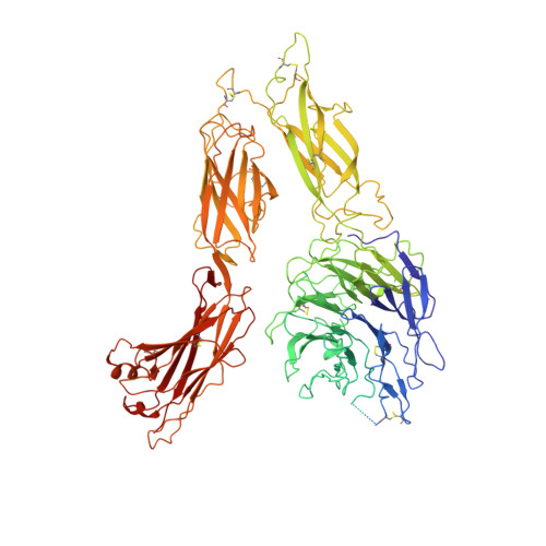
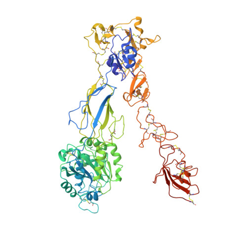
Structure of an integrin with an alphaI domain, complement receptor type 4.

Xie, C.Zhu, J.Chen, X.Mi, L.Nishida, N.Springer, T.A.

(2010) EMBO J 29: 666-679

  • DOI: https://doi.org/10.1038/emboj.2009.367
  • Primary Citation of Related Structures:  
    3K6S, 3K71, 3K72

  • PubMed Abstract: 
  • We report the structure of an integrin with an alphaI domain, alpha(X)beta(2), the complement receptor type 4. It was earlier expected that a fixed orientation between the alphaI domain and the beta-propeller domain in which it is inserted would be required for allosteric signal transmission ...

    We report the structure of an integrin with an alphaI domain, alpha(X)beta(2), the complement receptor type 4. It was earlier expected that a fixed orientation between the alphaI domain and the beta-propeller domain in which it is inserted would be required for allosteric signal transmission. However, the alphaI domain is highly flexible, enabling two betaI domain conformational states to couple to three alphaI domain states, and greater accessibility for ligand recognition. Although alpha(X)beta(2) is bent similarly to integrins that lack alphaI domains, the terminal domains of the alpha- and beta-legs, calf-2 and beta-tail, are oriented differently than in alphaI-less integrins. Linkers extending to the transmembrane domains are unstructured. Previous mutations in the beta(2)-tail domain support the importance of extension, rather than a deadbolt, in integrin activation. The locations of further activating mutations and antibody epitopes show the critical role of extension, and conversion from the closed to the open headpiece conformation, in integrin activation. Differences among 10 molecules in crystal lattices provide unprecedented information on interdomain flexibility important for modelling integrin extension and activation.


    Organizational Affiliation

    Department of Pathology, Harvard Medical School, Immune Disease Institute and Children's Hospital, Boston, MA 02115, USA.


Macromolecules
Find similar proteins by:  (by identity cutoff)  |  3D Structure
Entity ID: 1
MoleculeChainsSequence LengthOrganismDetailsImage
Integrin alpha-XA, C, E, G1,095Homo sapiensMutation(s): 0 
Gene Names: CD11CITGAX
UniProt & NIH Common Fund Data Resources
Find proteins for P20702 (Homo sapiens)
Explore P20702 
Go to UniProtKB:  P20702
PHAROS:  P20702
Entity Groups  
Sequence Clusters30% Identity50% Identity70% Identity90% Identity95% Identity100% Identity
UniProt GroupP20702
Protein Feature View
Expand
  • Reference Sequence
Find similar proteins by:  (by identity cutoff)  |  3D Structure
Entity ID: 2
MoleculeChainsSequence LengthOrganismDetailsImage
Integrin beta-2B, D, F, H687Homo sapiensMutation(s): 0 
Gene Names: CD18ITGB2MFI7
UniProt & NIH Common Fund Data Resources
Find proteins for P05107 (Homo sapiens)
Explore P05107 
Go to UniProtKB:  P05107
PHAROS:  P05107
Entity Groups  
Sequence Clusters30% Identity50% Identity70% Identity90% Identity95% Identity100% Identity
UniProt GroupP05107
Protein Feature View
Expand
  • Reference Sequence
Oligosaccharides

Help

Entity ID: 3
MoleculeChainsChain Length2D DiagramGlycosylation3D Interactions
2-acetamido-2-deoxy-beta-D-glucopyranose-(1-4)-2-acetamido-2-deoxy-beta-D-glucopyranoseI, M, N, P, S 2N-Glycosylation Oligosaccharides Interaction
Glycosylation Resources
GlyTouCan:  G42666HT
GlyCosmos:  G42666HT
GlyGen:  G42666HT
Entity ID: 4
MoleculeChainsChain Length2D DiagramGlycosylation3D Interactions
alpha-D-mannopyranose-(1-4)-2-acetamido-2-deoxy-beta-D-glucopyranose-(1-4)-2-acetamido-2-deoxy-beta-D-glucopyranose
J, K, L, O, Q, R, U
J, K, L, O, Q, R, U
3N-Glycosylation Oligosaccharides Interaction
Glycosylation Resources
GlyTouCan:  G62182OO
GlyCosmos:  G62182OO
GlyGen:  G62182OO
Entity ID: 5
MoleculeChainsChain Length2D DiagramGlycosylation3D Interactions
alpha-D-mannopyranose-(1-6)-alpha-D-mannopyranose-(1-4)-2-acetamido-2-deoxy-beta-D-glucopyranose-(1-4)-2-acetamido-2-deoxy-beta-D-glucopyranoseT 4N-Glycosylation Oligosaccharides Interaction
Glycosylation Resources
GlyTouCan:  G04854NQ
GlyCosmos:  G04854NQ
GlyGen:  G04854NQ
Small Molecules
Ligands 3 Unique
IDChainsName / Formula / InChI Key2D Diagram3D Interactions
NAG
Query on NAG

Download Ideal Coordinates CCD File 
AA [auth B],
CA [auth C],
DA [auth C],
HA [auth D],
IA [auth D],
AA [auth B],
CA [auth C],
DA [auth C],
HA [auth D],
IA [auth D],
KA [auth E],
LA [auth E],
PA [auth F],
QA [auth F],
SA [auth G],
TA [auth G],
V [auth A],
YA [auth H],
Z [auth B],
ZA [auth H]
2-acetamido-2-deoxy-beta-D-glucopyranose
C8 H15 N O6
OVRNDRQMDRJTHS-FMDGEEDCSA-N
 Ligand Interaction
CA
Query on CA

Download Ideal Coordinates CCD File 
AB [auth H],
BA [auth B],
EA [auth C],
FA [auth C],
GA [auth C],
AB [auth H],
BA [auth B],
EA [auth C],
FA [auth C],
GA [auth C],
JA [auth D],
MA [auth E],
NA [auth E],
OA [auth E],
RA [auth F],
UA [auth G],
VA [auth G],
W [auth A],
WA [auth G],
X [auth A],
Y [auth A]
CALCIUM ION
Ca
BHPQYMZQTOCNFJ-UHFFFAOYSA-N
 Ligand Interaction
MG
Query on MG

Download Ideal Coordinates CCD File 
XA [auth G]MAGNESIUM ION
Mg
JLVVSXFLKOJNIY-UHFFFAOYSA-N
 Ligand Interaction
Experimental Data & Validation

Experimental Data

  • Method: X-RAY DIFFRACTION
  • Resolution: 3.95 Å
  • R-Value Free: 0.373 
  • R-Value Work: 0.350 
  • R-Value Observed: 0.350 
  • Space Group: P 21 21 21
Unit Cell:
Length ( Å )Angle ( ˚ )
a = 149.664α = 90
b = 165.745β = 90
c = 537.676γ = 90
Software Package:
Software NamePurpose
MAR345dtbdata collection
PHASERphasing
PHENIXrefinement
XDSdata reduction
XDSdata scaling

Structure Validation

View Full Validation Report


Ligand Structure Quality Assessment 


Entry History 

Deposition Data

Revision History  (Full details and data files)

  • Version 1.0: 2010-01-12
    Type: Initial release
  • Version 1.1: 2011-07-13
    Changes: Version format compliance
  • Version 2.0: 2020-07-29
    Type: Remediation
    Reason: Carbohydrate remediation
    Changes: Advisory, Atomic model, Data collection, Database references, Derived calculations, Structure summary