3K70 | pdb_00003k70

Crystal structure of the complete initiation complex of RecBCD


Experimental Data Snapshot

  • Method: X-RAY DIFFRACTION
  • Resolution: 3.59 Å
  • R-Value Free: 
    0.296 (Depositor), 0.280 (DCC) 
  • R-Value Work: 
    0.248 (Depositor), 0.237 (DCC) 
  • R-Value Observed: 
    0.248 (Depositor) 

Starting Model: experimental
View more details

wwPDB Validation 3D Report Full Report

Validation slider image for 3K70

This is version 1.2 of the entry. See complete history

Literature

DNA binding to RecD: role of the 1B domain in SF1B helicase activity.

Saikrishnan, K.Griffiths, S.P.Cook, N.Court, R.Wigley, D.B.

(2008) EMBO J 27: 2222-2229

  • DOI: https://doi.org/10.1038/emboj.2008.144
  • Primary Citation Related Structures: 
    3E1S, 3K70

  • PubMed Abstract: 

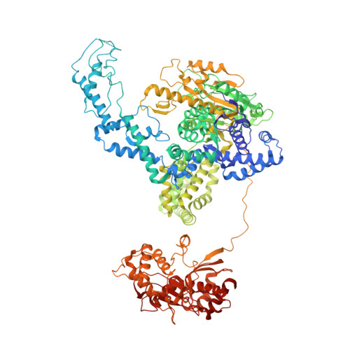
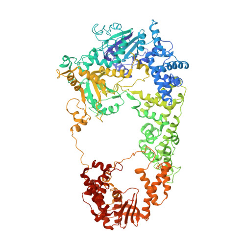
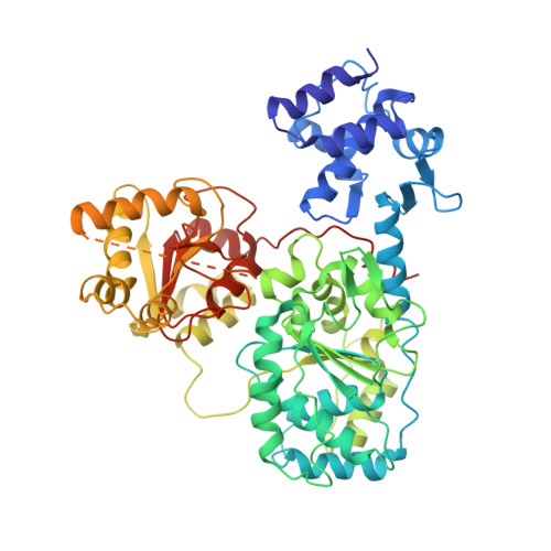
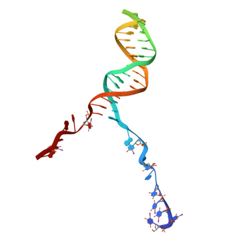
    The molecular mechanism of superfamily 1Balpha helicases remains unclear. We present here the crystal structure of the RecD2 helicase from Deinococcus radiodurans at 2.2-A resolution. The structure reveals the folds of the 1B and 2B domains of RecD that were poorly ordered in the structure of the Escherichia coli RecBCD enzyme complex reported previously. The 2B domain adopts an SH3 fold which, although common in eukaryotes, is extremely rare in bacterial systems. In addition, the D. radiodurans RecD2 structure has aided us in deciphering lower resolution (3.6 A) electron density maps for the E. coli RecBCD enzyme in complex with a long DNA substrate that interacts with the RecD subunit. Taken together, these structures indicated an important role for the 1B domain of RecD, a beta-hairpin that extends from the surface of the 1A domain and interacts with the DNA substrate. On the basis of these structural data, we designed a mutant RecD2 helicase that lacks this pin. The 'pin-less' mutant protein is a fully active ssDNA-dependent ATPase but totally lacks helicase activity.


  • Organizational Affiliation
    • Cancer Research UK Clare Hall Laboratories, The London Research Institute, Herts, UK.

Macromolecule Content 

  • Total Structure Weight: 693.49 kDa 
  • Atom Count: 46,932 
  • Modeled Residue Count: 5,738 
  • Deposited Residue Count: 5,922 
  • Unique protein chains: 3
  • Unique nucleic acid chains: 1

Macromolecules


Find similar proteins by:|  3D Structure
Entity ID: 1
MoleculeChains  Sequence LengthOrganismDetailsImage
Exodeoxyribonuclease V beta chainA [auth B],
D [auth E]
1,180Escherichia coli K-12Mutation(s): 0 
Gene Names: b2820JW2788recBrorA
EC: 3.1.11.5 (PDB Primary Data), 5.6.2.4 (UniProt)
UniProt
Find proteins for P08394 (Escherichia coli (strain K12))
Explore P08394 
Go to UniProtKB:  P08394
Entity Groups
Sequence Clusters30% Identity50% Identity70% Identity90% Identity95% Identity100% Identity
UniProt GroupP08394
Sequence Annotations
Expand
Reference Sequence
Find similar proteins by:|  3D Structure
Entity ID: 2
MoleculeChains  Sequence LengthOrganismDetailsImage
Exodeoxyribonuclease V gamma chainB [auth C],
E [auth F]
1,122Escherichia coli K-12Mutation(s): 0 
Gene Names: b2822JW2790recC
EC: 3.1.11.5
UniProt
Find proteins for P07648 (Escherichia coli (strain K12))
Explore P07648 
Go to UniProtKB:  P07648
Entity Groups
Sequence Clusters30% Identity50% Identity70% Identity90% Identity95% Identity100% Identity
UniProt GroupP07648
Sequence Annotations
Expand
Reference Sequence
Find similar proteins by:|  3D Structure
Entity ID: 3
MoleculeChains  Sequence LengthOrganismDetailsImage
Exodeoxyribonuclease V alpha chainC [auth D],
F [auth G]
608Escherichia coli K-12Mutation(s): 0 
Gene Names: b2819JW2787recD
EC: 3.1.11.5 (PDB Primary Data), 5.6.2.3 (UniProt)
UniProt
Find proteins for P04993 (Escherichia coli (strain K12))
Explore P04993 
Go to UniProtKB:  P04993
Entity Groups
Sequence Clusters30% Identity50% Identity70% Identity90% Identity95% Identity100% Identity
UniProt GroupP04993
Sequence Annotations
Expand
Reference Sequence
Find similar nucleic acids by:  (by identity cutoff) 
Entity ID: 4
MoleculeChains LengthOrganismImage
DNA (46-MER)G [auth X],
H [auth Y]
51N/A
Sequence Annotations
Expand
Reference Sequence

Experimental Data & Validation

Experimental Data

  • Method: X-RAY DIFFRACTION
  • Resolution: 3.59 Å
  • R-Value Free:  0.296 (Depositor), 0.280 (DCC) 
  • R-Value Work:  0.248 (Depositor), 0.237 (DCC) 
  • R-Value Observed: 0.248 (Depositor) 
Space Group: P 21 21 21
Unit Cell:
Length ( Å )Angle ( ˚ )
a = 133.8α = 90
b = 192.9β = 90
c = 334.8γ = 90
Software Package:
Software NamePurpose
ADSCdata collection
PHASERphasing
CNSrefinement
MOSFLMdata reduction
SCALAdata scaling

Structure Validation

View Full Validation Report


Entry History 

Deposition Data

Revision History  (Full details and data files)

  • Version 1.0: 2009-11-10
    Type: Initial release
  • Version 1.1: 2011-07-13
    Changes: Version format compliance
  • Version 1.2: 2023-09-06
    Changes: Data collection, Database references, Derived calculations, Refinement description