3K4V

New crystal form of HIV-1 Protease/Saquinavir structure reveals carbamylation of N-terminal proline


Experimental Data Snapshot

  • Method: X-RAY DIFFRACTION
  • Resolution: 1.39 Å
  • R-Value Free: 0.227 
  • R-Value Observed: 0.159 

wwPDB Validation   3D Report Full Report


Ligand Structure Quality Assessment 


This is version 1.6 of the entry. See complete history


Literature

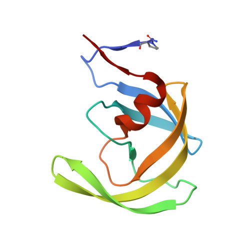
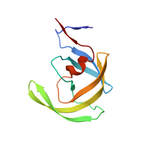
Carbamylation of N-terminal proline.

Olajuyigbe, F.M.Demitri, N.Ajele, J.O.Maurizio, E.Randaccio, L.Geremia, S.

(2010) ACS Med Chem Lett 1: 254-257

  • DOI: https://doi.org/10.1021/ml100046d
  • Primary Citation of Related Structures:  
    3K4V

  • PubMed Abstract: 

    Protein carbamylation is of great concern both in vivo and in vitro. Here, we report the first structural characterization of a protein carbamylated at the N-terminal proline. The unexpected carbamylation of the α-amino group of the least reactive codified amino acid has been detected in high-resolution electron density maps of a new crystal form of the HIV-1 protease/saquinavir complex. The carbamyl group is found coplanar to the proline ring with a trans conformation. The reaction of N-terminal with cyanate ion derived from the chaotropic agent urea was confirmed by mass spectra analysis on protease single crystals. Implications of carbamylation process in vitro and in vivo are discussed.


  • Organizational Affiliation

    Department of Chemical Sciences ; Department of Biochemistry, Federal University of Technology, 340001 Akure, Nigeria.


Macromolecules
Find similar proteins by:  (by identity cutoff)  |  3D Structure
Entity ID: 1
MoleculeChains Sequence LengthOrganismDetailsImage
HIV-1 Protease
A, B
100Human immunodeficiency virus type 1 BH10Mutation(s): 5 
Gene Names: gag-pol
EC: 3.4.23.16
UniProt
Find proteins for P03366 (Human immunodeficiency virus type 1 group M subtype B (isolate BH10))
Explore P03366 
Go to UniProtKB:  P03366
Entity Groups  
Sequence Clusters30% Identity50% Identity70% Identity90% Identity95% Identity100% Identity
UniProt GroupP03366
Sequence Annotations
Expand
  • Reference Sequence
Find similar proteins by:  (by identity cutoff)  |  3D Structure
Entity ID: 2
MoleculeChains Sequence LengthOrganismDetailsImage
HIV-1 Protease
C, D
99Human immunodeficiency virus type 1 BH10Mutation(s): 5 
Gene Names: gag-pol
EC: 3.4.23.16
UniProt
Find proteins for P03366 (Human immunodeficiency virus type 1 group M subtype B (isolate BH10))
Explore P03366 
Go to UniProtKB:  P03366
Entity Groups  
Sequence Clusters30% Identity50% Identity70% Identity90% Identity95% Identity100% Identity
UniProt GroupP03366
Sequence Annotations
Expand
  • Reference Sequence
Small Molecules
Ligands 3 Unique
IDChains Name / Formula / InChI Key2D Diagram3D Interactions
ROC
Query on ROC

Download Ideal Coordinates CCD File 
G [auth B],
J [auth D]
(2S)-N-[(2S,3R)-4-[(2S,3S,4aS,8aS)-3-(tert-butylcarbamoyl)-3,4,4a,5,6,7,8,8a-octahydro-1H-isoquinolin-2-yl]-3-hydroxy-1 -phenyl-butan-2-yl]-2-(quinolin-2-ylcarbonylamino)butanediamide
C38 H50 N6 O5
QWAXKHKRTORLEM-UGJKXSETSA-N
DMS
Query on DMS

Download Ideal Coordinates CCD File 
E [auth A],
F [auth A],
I [auth C],
K [auth D]
DIMETHYL SULFOXIDE
C2 H6 O S
IAZDPXIOMUYVGZ-UHFFFAOYSA-N
CL
Query on CL

Download Ideal Coordinates CCD File 
H [auth B]CHLORIDE ION
Cl
VEXZGXHMUGYJMC-UHFFFAOYSA-M
Binding Affinity Annotations 
IDSourceBinding Affinity
ROC BindingDB:  3K4V Ki: min: 0.04, max: 138 (nM) from 29 assay(s)
Kd: min: 0.31, max: 67 (nM) from 6 assay(s)
IC50: min: 0.2, max: 270 (nM) from 16 assay(s)
-TΔS: min: -8.03e+1, max: -2.80e+1 (kJ/mol) from 47 assay(s)
ΔH: min: -3.18e+1, max: 43.47 (kJ/mol) from 47 assay(s)
ΔG: min: -6.23e+1, max: -3.55e+1 (kJ/mol) from 44 assay(s)
Biologically Interesting Molecules (External Reference) 1 Unique
Experimental Data & Validation

Experimental Data

  • Method: X-RAY DIFFRACTION
  • Resolution: 1.39 Å
  • R-Value Free: 0.227 
  • R-Value Observed: 0.159 
  • Space Group: P 1 21 1
Unit Cell:
Length ( Å )Angle ( ˚ )
a = 51.205α = 90
b = 62.673β = 98.42
c = 59.076γ = 90
Software Package:
Software NamePurpose
HKL-2000data collection
AMoREphasing
SHELXL-97refinement
MOSFLMdata reduction
SCALAdata scaling

Structure Validation

View Full Validation Report


Ligand Structure Quality Assessment 


Entry History 

Deposition Data

Revision History  (Full details and data files)

  • Version 1.0: 2010-06-09
    Type: Initial release
  • Version 1.1: 2011-07-13
    Changes: Atomic model, Database references, Derived calculations, Non-polymer description, Structure summary, Version format compliance
  • Version 1.2: 2011-12-07
    Changes: Database references
  • Version 1.3: 2012-12-12
    Changes: Other
  • Version 1.4: 2014-09-24
    Changes: Database references
  • Version 1.5: 2021-10-13
    Changes: Database references, Derived calculations, Structure summary
  • Version 1.6: 2023-09-06
    Changes: Data collection, Refinement description