


Structure of the pterin-binding domain MeTr of 5-methyltetrahydrofolate-homocysteine methyltransferase from Bacteroides thetaiotaomicron
Cuff, M.E., Li, H., Cobb, G., Joachimiak, A.To be published.
Experimental Data Snapshot
Entity ID: 1 | |||||
|---|---|---|---|---|---|
| Molecule | Chains | Sequence Length | Organism | Details | Image |
| 5-methyltetrahydrofolate-homocysteine methyltransferase | 300 | Bacteroides thetaiotaomicron | Mutation(s): 0 Gene Names: BT_0180 EC: 2.1.1.13 | 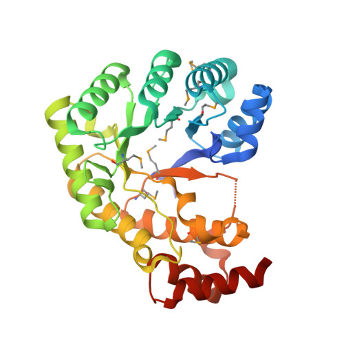 | |
UniProt | |||||
Entity Groups | |||||
| Sequence Clusters | 30% Identity50% Identity70% Identity90% Identity95% Identity100% Identity | ||||
| UniProt Group | Q8ABD0 | ||||
Sequence AnnotationsExpand | |||||
Reference Sequence | |||||
| Ligands 4 Unique | |||||
|---|---|---|---|---|---|
| ID | Chains | Name / Formula / InChI Key | 2D Diagram | 3D Interactions | |
| THH Download:Ideal Coordinates CCD File | E [auth A], M [auth B], T [auth C] | N-[4-({[(6S)-2-AMINO-4-HYDROXY-5-METHYL-5,6,7,8-TETRAHYDROPTERIDIN-6-YL]METHYL}AMINO)BENZOYL]-L-GLUTAMIC ACID C20 H25 N7 O6 ZNOVTXRBGFNYRX-STQMWFEESA-N |  | ||
| GOL Download:Ideal Coordinates CCD File | D [auth A] F [auth A] I [auth A] K [auth B] L [auth B] | GLYCEROL C3 H8 O3 PEDCQBHIVMGVHV-UHFFFAOYSA-N |  | ||
| K Download:Ideal Coordinates CCD File | G [auth A], H [auth A] | POTASSIUM ION K NPYPAHLBTDXSSS-UHFFFAOYSA-N |  | ||
| NA Download:Ideal Coordinates CCD File | J [auth A], N [auth B], Q [auth B], U [auth C], W [auth C] | SODIUM ION Na FKNQFGJONOIPTF-UHFFFAOYSA-N |  | ||
| Modified Residues 1 Unique | |||||
|---|---|---|---|---|---|
| ID | Chains | Type | Formula | 2D Diagram | Parent |
| MSE Query on MSE | A, B, C | L-PEPTIDE LINKING | C5 H11 N O2 Se |  | MET |
| Length ( Å ) | Angle ( ˚ ) |
|---|---|
| a = 137.652 | α = 90 |
| b = 79.992 | β = 90.12 |
| c = 127.068 | γ = 90 |
| Software Name | Purpose |
|---|---|
| DENZO | data reduction |
| SCALEPACK | data scaling |
| MLPHARE | phasing |
| DM | phasing |
| REFMAC | refinement |
| PDB_EXTRACT | data extraction |
| SBC-Collect | data collection |
| HKL-3000 | data reduction |
| HKL-3000 | data scaling |
| HKL-3000 | phasing |
| SHELXD | phasing |
| SHELXE | model building |
| SOLVE | phasing |
| RESOLVE | phasing |
| ARP/wARP | model building |
| CCP4 | phasing |
| O | model building |
| Coot | model building |


