3JWO

Structure of HIV-1 gp120 with gp41-Interactive Region: Layered Architecture and Basis of Conformational Mobility


Experimental Data Snapshot

  • Method: X-RAY DIFFRACTION
  • Resolution: 3.51 Å
  • R-Value Free: 0.273 
  • R-Value Work: 0.257 
  • R-Value Observed: 0.258 

wwPDB Validation   3D Report Full Report


Ligand Structure Quality Assessment 


This is version 2.1 of the entry. See complete history


Literature

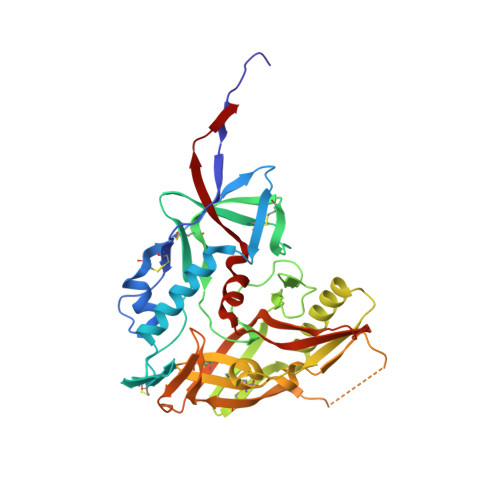
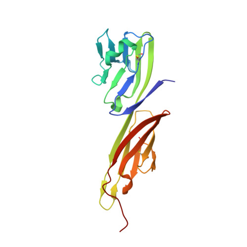
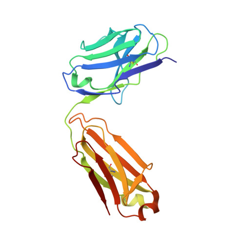
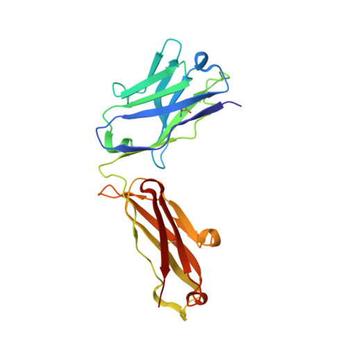
Structure of HIV-1 gp120 with gp41-interactive region reveals layered envelope architecture and basis of conformational mobility.

Pancera, M.Majeed, S.Ban, Y.E.Chen, L.Huang, C.C.Kong, L.Kwon, Y.D.Stuckey, J.Zhou, T.Robinson, J.E.Schief, W.R.Sodroski, J.Wyatt, R.Kwong, P.D.

(2010) Proc Natl Acad Sci U S A 107: 1166-1171

  • DOI: https://doi.org/10.1073/pnas.0911004107
  • Primary Citation of Related Structures:  
    3JWD, 3JWO

  • PubMed Abstract: 

    The viral spike of HIV-1 is composed of three gp120 envelope glycoproteins attached noncovalently to three gp41 transmembrane molecules. Viral entry is initiated by binding to the CD4 receptor on the cell surface, which induces large conformational changes in gp120. These changes not only provide a model for receptor-triggered entry, but affect spike sensitivity to drug- and antibody-mediated neutralization. Although some of the details of the CD4-induced conformational change have been visualized by crystal structures and cryoelectron tomograms, the critical gp41-interactive region of gp120 was missing from previous atomic-level characterizations. Here we determine the crystal structure of an HIV-1 gp120 core with intact gp41-interactive region in its CD4-bound state, compare this structure to unliganded and antibody-bound forms to identify structurally invariant and plastic components, and use ligand-oriented cryoelectron tomograms to define component mobility in the viral spike context. Newly defined gp120 elements proximal to the gp41 interface complete a 7-stranded beta-sandwich, which appeared invariant in conformation. Loop excursions emanating from the sandwich form three topologically separate--and structurally plastic--layers, topped off by the highly glycosylated gp120 outer domain. Crystal structures, cryoelectron tomograms, and interlayer chemistry were consistent with a mechanism in which the layers act as a shape-changing spacer, facilitating movement between outer domain and gp41-associated beta-sandwich and providing for conformational diversity used in immune evasion. A "layered" gp120 architecture thus allows movement among alternative glycoprotein conformations required for virus entry and immune evasion, whereas a beta-sandwich clamp maintains gp120-gp41 interaction and regulates gp41 transitions.


  • Organizational Affiliation

    Vaccine Research Center, National Institute of Allergy and Infectious Diseases, National Institutes of Health, Bethesda, MD 20892, USA.


Macromolecules
Find similar proteins by:  (by identity cutoff)  |  3D Structure
Entity ID: 1
MoleculeChains Sequence LengthOrganismDetailsImage
HIV-1 GP120 ENVELOPE GLYCOPROTEIN357Homo sapiensMutation(s): 0 
Entity Groups  
Sequence Clusters30% Identity50% Identity70% Identity90% Identity95% Identity100% Identity
Sequence Annotations
Expand
  • Reference Sequence
Find similar proteins by:  (by identity cutoff)  |  3D Structure
Entity ID: 2
MoleculeChains Sequence LengthOrganismDetailsImage
T-cell surface glycoprotein CD4B [auth C]184Homo sapiensMutation(s): 0 
Gene Names: CD4
UniProt & NIH Common Fund Data Resources
Find proteins for P01730 (Homo sapiens)
Explore P01730 
Go to UniProtKB:  P01730
PHAROS:  P01730
GTEx:  ENSG00000010610 
Entity Groups  
Sequence Clusters30% Identity50% Identity70% Identity90% Identity95% Identity100% Identity
UniProt GroupP01730
Sequence Annotations
Expand
  • Reference Sequence
Find similar proteins by:  (by identity cutoff)  |  3D Structure
Entity ID: 3
MoleculeChains Sequence LengthOrganismDetailsImage
FAB 48D LIGHT CHAINC [auth L]209Homo sapiensMutation(s): 0 
Entity Groups  
Sequence Clusters30% Identity50% Identity70% Identity90% Identity95% Identity100% Identity
Sequence Annotations
Expand
  • Reference Sequence
Find similar proteins by:  (by identity cutoff)  |  3D Structure
Entity ID: 4
MoleculeChains Sequence LengthOrganismDetailsImage
FAB 48D Heavy CHAIND [auth H]220Homo sapiensMutation(s): 0 
Entity Groups  
Sequence Clusters30% Identity50% Identity70% Identity90% Identity95% Identity100% Identity
Sequence Annotations
Expand
  • Reference Sequence
Oligosaccharides

Help

Entity ID: 5
MoleculeChains Length2D Diagram Glycosylation3D Interactions
beta-L-fucopyranose-(1-6)-2-acetamido-2-deoxy-beta-D-glucopyranoseE [auth B]2N-Glycosylation
Glycosylation Resources
GlyTouCan:  G61843VN
GlyCosmos:  G61843VN
GlyGen:  G61843VN
Experimental Data & Validation

Experimental Data

  • Method: X-RAY DIFFRACTION
  • Resolution: 3.51 Å
  • R-Value Free: 0.273 
  • R-Value Work: 0.257 
  • R-Value Observed: 0.258 
  • Space Group: P 41 21 2
Unit Cell:
Length ( Å )Angle ( ˚ )
a = 190α = 90
b = 190β = 90
c = 103.302γ = 90
Software Package:
Software NamePurpose
DENZOdata reduction
SCALEPACKdata scaling
AMoREphasing
PHENIXrefinement
PDB_EXTRACTdata extraction
HKL-2000data collection

Structure Validation

View Full Validation Report


Ligand Structure Quality Assessment 


Entry History 

Deposition Data

Revision History  (Full details and data files)

  • Version 1.0: 2009-12-29
    Type: Initial release
  • Version 1.1: 2011-07-13
    Changes: Non-polymer description, Version format compliance
  • Version 1.2: 2017-11-01
    Changes: Refinement description
  • Version 2.0: 2020-07-29
    Type: Remediation
    Reason: Carbohydrate remediation
    Changes: Advisory, Atomic model, Data collection, Database references, Derived calculations, Structure summary
  • Version 2.1: 2023-09-06
    Changes: Data collection, Database references, Refinement description, Structure summary