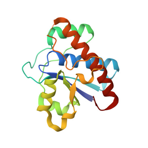
3JVI

Product state mimic crystal structure of protein tyrosine phosphatase from Entamoeba histolytica


Experimental Data Snapshot

  • Method: X-RAY DIFFRACTION
  • Resolution: 1.80 Å
  • R-Value Free: 0.249 
  • R-Value Work: 0.204 
  • R-Value Observed: 0.206 

Starting Model: experimental
View more details

wwPDB Validation   3D Report Full Report


This is version 1.5 of the entry. See complete history


Literature

Crystal structure and putative substrate identification for the Entamoeba histolytica low molecular weight tyrosine phosphatase.

Linford, A.S.Jiang, N.M.Edwards, T.E.Sherman, N.E.Van Voorhis, W.C.Stewart, L.J.Myler, P.J.Staker, B.L.Petri, W.A.

(2014) Mol Biochem Parasitol 193: 33-44

  • DOI: https://doi.org/10.1016/j.molbiopara.2014.01.003
  • Primary Citation of Related Structures:  
    3IDO, 3ILY, 3JS5, 3JVI

  • PubMed Abstract: 

    Entamoeba histolytica is a eukaryotic intestinal parasite of humans, and is endemic in developing countries. We have characterized the E. histolytica putative low molecular weight protein tyrosine phosphatase (LMW-PTP). The structure for this amebic tyrosine phosphatase was solved, showing the ligand-induced conformational changes necessary for binding of substrate. In amebae, it was expressed at low but detectable levels as detected by immunoprecipitation followed by immunoblotting. A mutant LMW-PTP protein in which the catalytic cysteine in the active site was replaced with a serine lacked phosphatase activity, and was used to identify a number of trapped putative substrate proteins via mass spectrometry analysis. Seven of these putative substrate protein genes were cloned with an epitope tag and overexpressed in amebae. Five of these seven putative substrate proteins were demonstrated to interact specifically with the mutant LMW-PTP. This is the first biochemical study of a small tyrosine phosphatase in Entamoeba, and sets the stage for understanding its role in amebic biology and pathogenesis.


  • Organizational Affiliation

    Department of Biochemistry and Molecular Genetics, University of Virginia Health System, Charlottesville, VA 22908, USA. Electronic address: asl2c@virginia.edu.


Macromolecules
Find similar proteins by:  (by identity cutoff)  |  3D Structure
Entity ID: 1
MoleculeChains Sequence LengthOrganismDetailsImage
Protein tyrosine phosphatase161Entamoeba histolytica HM-1:IMSSMutation(s): 0 
Gene Names: EHI_153650
UniProt
Find proteins for C4LSE7 (Entamoeba histolytica (strain ATCC 30459 / HM-1:IMSS / ABRM))
Explore C4LSE7 
Go to UniProtKB:  C4LSE7
Entity Groups  
Sequence Clusters30% Identity50% Identity70% Identity90% Identity95% Identity100% Identity
UniProt GroupC4LSE7
Sequence Annotations
Expand
  • Reference Sequence
Small Molecules
Ligands 1 Unique
IDChains Name / Formula / InChI Key2D Diagram3D Interactions
SO4
Query on SO4

Download Ideal Coordinates CCD File 
B [auth A]SULFATE ION
O4 S
QAOWNCQODCNURD-UHFFFAOYSA-L
Experimental Data & Validation

Experimental Data

  • Method: X-RAY DIFFRACTION
  • Resolution: 1.80 Å
  • R-Value Free: 0.249 
  • R-Value Work: 0.204 
  • R-Value Observed: 0.206 
  • Space Group: P 43 21 2
Unit Cell:
Length ( Å )Angle ( ˚ )
a = 44.995α = 90
b = 44.995β = 90
c = 133.316γ = 90
Software Package:
Software NamePurpose
PHASERphasing
REFMACrefinement
PDB_EXTRACTdata extraction
HKL-2000data reduction
HKL-2000data scaling

Structure Validation

View Full Validation Report


Entry History 

Deposition Data

Revision History  (Full details and data files)

  • Version 1.0: 2009-09-22
    Type: Initial release
  • Version 1.1: 2011-07-13
    Changes: Advisory, Refinement description, Source and taxonomy, Version format compliance
  • Version 1.2: 2014-03-12
    Changes: Database references
  • Version 1.3: 2014-04-09
    Changes: Database references
  • Version 1.4: 2017-11-01
    Changes: Refinement description
  • Version 1.5: 2023-09-06
    Changes: Data collection, Database references, Derived calculations, Refinement description