3JV2

Crystal Structure of B. subtilis SecA with bound peptide


Experimental Data Snapshot

  • Method: X-RAY DIFFRACTION
  • Resolution: 2.50 Å
  • R-Value Free: 0.258 
  • R-Value Work: 0.208 

wwPDB Validation   3D Report Full Report


Ligand Structure Quality Assessment 


This is version 1.2 of the entry. See complete history


Literature

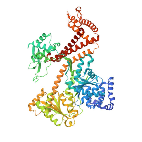
Conformational flexibility and peptide interaction of the translocation ATPase SecA.

Zimmer, J.Rapoport, T.A.

(2009) J Mol Biol 394: 606-612

  • DOI: https://doi.org/10.1016/j.jmb.2009.10.024
  • Primary Citation of Related Structures:  
    3JUX, 3JV2

  • PubMed Abstract: 

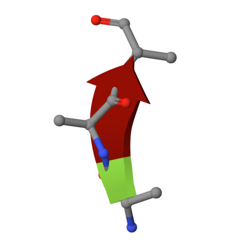
    The SecA ATPase forms a functional complex with the protein-conducting SecY channel to translocate polypeptides across the bacterial cell membrane. SecA recognizes the translocation substrate and catalyzes its unidirectional movement through the SecY channel. The recent crystal structure of the Thermotoga maritima SecA-SecYEG complex shows the ATPase in a conformation where the nucleotide-binding domains (NBDs) have closed around a bound ADP-BeFx complex and SecA's polypeptide-binding clamp is shut. Here, we present the crystal structure of T. maritima SecA in isolation, determined in its ADP-bound form at 3.1 A resolution. SecA alone has a drastically different conformation in which the nucleotide-binding pocket between NBD1 and NBD2 is open and the preprotein cross-linking domain has rotated away from both NBDs, thereby opening the polypeptide-binding clamp. To investigate how this clamp binds polypeptide substrates, we also determined a structure of Bacillus subtilis SecA in complex with a peptide at 2.5 A resolution. This structure shows that the peptide augments the highly conserved beta-sheet at the back of the clamp. Taken together, these structures suggest a mechanism by which ATP hydrolysis can lead to polypeptide translocation.


  • Organizational Affiliation

    Howard Hughes Medical Institute and Department of Cell Biology, Harvard Medical School, 240 Longwood Avenue, Boston, MA 02115, USA.


Macromolecules
Find similar proteins by:  (by identity cutoff)  |  3D Structure
Entity ID: 1
MoleculeChains Sequence LengthOrganismDetailsImage
Protein translocase subunit SecA
A, B
783Bacillus subtilisMutation(s): 0 
Gene Names: secA
UniProt
Find proteins for P28366 (Bacillus subtilis (strain 168))
Explore P28366 
Go to UniProtKB:  P28366
Entity Groups  
Sequence Clusters30% Identity50% Identity70% Identity90% Identity95% Identity100% Identity
UniProt GroupP28366
Sequence Annotations
Expand
  • Reference Sequence

Find similar proteins by:  Sequence   |   3D Structure  

Entity ID: 2
MoleculeChains Sequence LengthOrganismDetailsImage
peptide
C, D
3N/AMutation(s): 0 
Sequence Annotations
Expand
  • Reference Sequence
Experimental Data & Validation

Experimental Data

  • Method: X-RAY DIFFRACTION
  • Resolution: 2.50 Å
  • R-Value Free: 0.258 
  • R-Value Work: 0.208 
  • Space Group: P 41
Unit Cell:
Length ( Å )Angle ( ˚ )
a = 106.724α = 90
b = 106.724β = 90
c = 175.605γ = 90
Software Package:
Software NamePurpose
HKL-2000data collection
PHASERphasing
REFMACrefinement
HKL-2000data reduction
HKL-2000data scaling

Structure Validation

View Full Validation Report


Ligand Structure Quality Assessment 


Entry History 

Deposition Data

  • Released Date: 2009-11-24 
  • Deposition Author(s): Zimmer, J.

Revision History  (Full details and data files)

  • Version 1.0: 2009-11-24
    Type: Initial release
  • Version 1.1: 2011-07-13
    Changes: Version format compliance
  • Version 1.2: 2023-09-06
    Changes: Data collection, Database references, Derived calculations, Refinement description, Source and taxonomy, Structure summary