3JQZ | pdb_00003jqz

Crystal Structure of Human serum albumin complexed with Lidocaine


Experimental Data Snapshot

  • Method: X-RAY DIFFRACTION
  • Resolution: 3.30 Å
  • R-Value Free: 
    0.268 (Depositor), 0.263 (DCC) 
  • R-Value Work: 
    0.220 (Depositor), 0.213 (DCC) 
  • R-Value Observed: 
    0.223 (Depositor) 

Starting Model: experimental
View more details

wwPDB Validation 3D Report Full Report

Validation slider image for 3JQZ

Ligand Structure Quality Assessment 


This is version 1.4 of the entry. See complete history

Literature

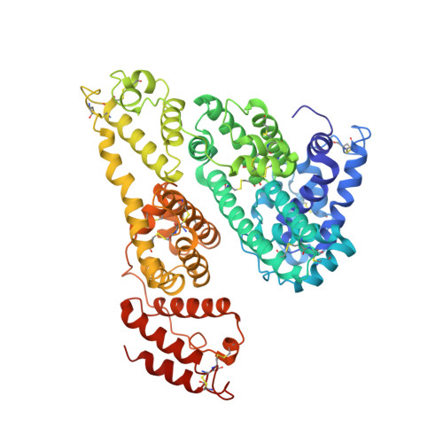
Crystallographic analysis reveals a unique lidocaine binding site on human serum albumin.

Hein, K.L.Kragh-Hansen, U.Morth, J.P.Jeppesen, M.D.Otzen, D.Moller, J.V.Nissen, P.

(2010) J Struct Biol 

  • DOI: https://doi.org/10.1016/j.jsb.2010.03.014
  • Primary Citation Related Structures: 
    3JQZ, 3JRY

  • PubMed Abstract: 

    Human serum albumin (HSA), the major protein component in blood plasma and in extravascular spaces, is known to participate in the binding and transport of a variety of endogenous and exogenous organic compounds with anionic or electronegative features. We here report on the 3.3A resolution crystal structure of HSA complexed with the cationic, and widely used, anesthetic lidocaine. We find that lidocaine and HSA co-crystallise as a dimer in the unusual space group I4(1). The dimer consists of one HSA molecule without ligand and one HSA molecule with a single, bound lidocaine. HSA is a heart-shaped protein composed of three homologous helical domains (I-III), which can be subdivided into two subdomains (A and B), and lidocaine binds to a unique site formed by residues from subdomain IB facing the central, interdomain crevice. In the crystal, binding seems to introduce only local conformational changes in the protein. According to intrinsic fluorescence experiments with aqueous HSA binding results in widespread conformational changes involving Trp214 in subdomain IIA. Results obtained with equilibrium dialysis and isothermal titration calorimetry show that lidocaine binding is of a low affinity and occurs at one discrete binding site in accordance with the X-ray data. Another crystal form of ligand-free HSA obtained in the presence of ammonium sulphate was determined at 2.3A resolution revealing a sulphate ion accepting cavity at the surface of subdomain IIIA. The present results contribute to a further characterisation of the exceptional binding properties of HSA.


  • Organizational Affiliation
    • Department of Molecular Biology, University of Aarhus, Gustav Wieds Vej 10C, DK-8000 Arhus C, Denmark.

Macromolecule Content 

  • Total Structure Weight: 133.38 kDa 
  • Atom Count: 9,279 
  • Modeled Residue Count: 1,164 
  • Deposited Residue Count: 1,170 
  • Unique protein chains: 1

Macromolecules

Find similar proteins by:|  3D Structure
Entity ID: 1
MoleculeChains  Sequence LengthOrganismDetailsImage
Serum albumin
A, B
585Homo sapiensMutation(s): 0 
Gene Names: ALBGIG20GIG42PRO0903PRO1708PRO2044PRO2619PRO2675UNQ696/PRO1341
UniProt & NIH Common Fund Data Resources
Find proteins for P02768 (Homo sapiens)
Explore P02768 
Go to UniProtKB:  P02768
PHAROS:  P02768
GTEx:  ENSG00000163631 
Entity Groups
Sequence Clusters30% Identity50% Identity70% Identity90% Identity95% Identity100% Identity
UniProt GroupP02768
Sequence Annotations
Expand
Reference Sequence

Small Molecules

Ligands 1 Unique
IDChains Name / Formula / InChI Key2D Diagram3D Interactions
LQZ

Query on LQZ


Download:Ideal Coordinates CCD File
C [auth A]2-(diethylamino)-N-(2,6-dimethylphenyl)ethanamide
C14 H22 N2 O
NNJVILVZKWQKPM-UHFFFAOYSA-N

Experimental Data & Validation

Experimental Data

  • Method: X-RAY DIFFRACTION
  • Resolution: 3.30 Å
  • R-Value Free:  0.268 (Depositor), 0.263 (DCC) 
  • R-Value Work:  0.220 (Depositor), 0.213 (DCC) 
  • R-Value Observed: 0.223 (Depositor) 
Space Group: I 41
Unit Cell:
Length ( Å )Angle ( ˚ )
a = 168.476α = 90
b = 168.476β = 90
c = 97.02γ = 90
Software Package:
Software NamePurpose
RemDAqdata collection
PHASERphasing
PHENIXrefinement
XDSdata reduction
XDSdata scaling

Structure Validation

View Full Validation Report


Ligand Structure Quality Assessment 


Entry History 

Deposition Data

Revision History  (Full details and data files)

  • Version 1.0: 2010-04-21
    Type: Initial release
  • Version 1.1: 2011-07-13
    Changes: Version format compliance
  • Version 1.2: 2017-11-01
    Changes: Refinement description
  • Version 1.3: 2023-11-01
    Changes: Data collection, Database references, Derived calculations, Refinement description
  • Version 1.4: 2024-11-20
    Changes: Structure summary