3JBX | pdb_00003jbx

Cryo-electron microscopy structure of RAG Signal End Complex (C2 symmetry)


Experimental Data Snapshot

  • Method: ELECTRON MICROSCOPY
  • Resolution: 3.40 Å
  • Aggregation State: PARTICLE 
  • Reconstruction Method: SINGLE PARTICLE 

wwPDB Validation 3D Report Full Report

Validation slider image for 3JBX

This is version 1.2 of the entry. See complete history

Literature

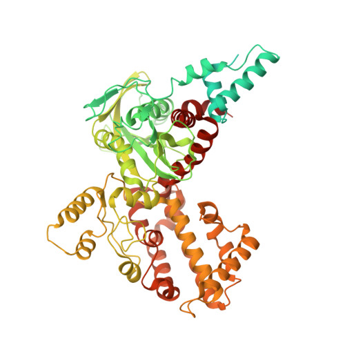
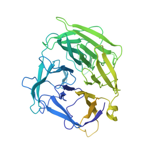
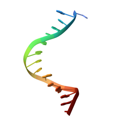
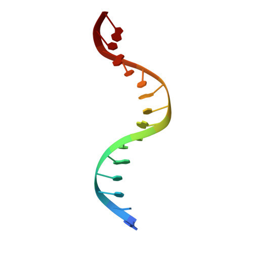
Molecular Mechanism of V(D)J Recombination from Synaptic RAG1-RAG2 Complex Structures.

Ru, H.Chambers, M.G.Fu, T.M.Tong, A.B.Liao, M.Wu, H.

(2015) Cell 163: 1138-1152

  • DOI: https://doi.org/10.1016/j.cell.2015.10.055
  • Primary Citation Related Structures: 
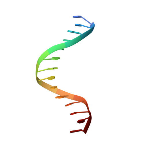
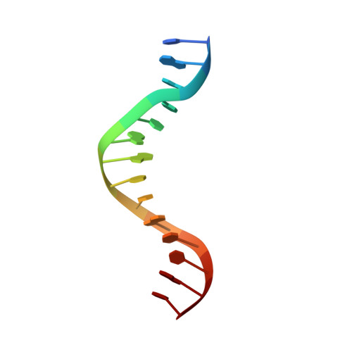
    3JBW, 3JBX, 3JBY

  • PubMed Abstract: 

    Diverse repertoires of antigen-receptor genes that result from combinatorial splicing of coding segments by V(D)J recombination are hallmarks of vertebrate immunity. The (RAG1-RAG2)2 recombinase (RAG) recognizes recombination signal sequences (RSSs) containing a heptamer, a spacer of 12 or 23 base pairs, and a nonamer (12-RSS or 23-RSS) and introduces precise breaks at RSS-coding segment junctions. RAG forms synaptic complexes only with one 12-RSS and one 23-RSS, a dogma known as the 12/23 rule that governs the recombination fidelity. We report cryo-electron microscopy structures of synaptic RAG complexes at up to 3.4 Å resolution, which reveal a closed conformation with base flipping and base-specific recognition of RSSs. Distortion at RSS-coding segment junctions and base flipping in coding segments uncover the two-metal-ion catalytic mechanism. Induced asymmetry involving tilting of the nonamer-binding domain dimer of RAG1 upon binding of HMGB1-bent 12-RSS or 23-RSS underlies the molecular mechanism for the 12/23 rule.


  • Organizational Affiliation
    • Department of Biological Chemistry and Molecular Pharmacology, Harvard Medical School, Boston, MA 02115, USA; Program in Cellular and Molecular Medicine, Boston Children's Hospital, Boston, MA 02115, USA.

Macromolecule Content 

  • Total Structure Weight: 329.95 kDa 
  • Atom Count: 16,670 
  • Modeled Residue Count: 1,918 
  • Deposited Residue Count: 2,710 
  • Unique protein chains: 2
  • Unique nucleic acid chains: 4

Macromolecules


Find similar proteins by:|  3D Structure
Entity ID: 1
MoleculeChains  Sequence LengthOrganismDetailsImage
V(D)J recombination-activating protein 1
A, C
764Danio rerioMutation(s): 0 
Gene Names: rag1
EC: 3.1 (PDB Primary Data), 6.3.2 (PDB Primary Data), 2.3.2.27 (UniProt)
UniProt
Find proteins for O13033 (Danio rerio)
Explore O13033 
Go to UniProtKB:  O13033
Entity Groups
Sequence Clusters30% Identity50% Identity70% Identity90% Identity95% Identity100% Identity
UniProt GroupO13033
Sequence Annotations
Expand
Reference Sequence
Find similar proteins by:|  3D Structure
Entity ID: 2
MoleculeChains  Sequence LengthOrganismDetailsImage
V(D)J recombination-activating protein 2
B, D
533Danio rerioMutation(s): 0 
Gene Names: rag2rag-2
UniProt
Find proteins for O13034 (Danio rerio)
Explore O13034 
Go to UniProtKB:  O13034
Entity Groups
Sequence Clusters30% Identity50% Identity70% Identity90% Identity95% Identity100% Identity
UniProt GroupO13034
Sequence Annotations
Expand
Reference Sequence
Find similar nucleic acids by:  Sequence
Entity ID: 3
MoleculeChains LengthOrganismImage
5'-D(*CP*AP*CP*AP*GP*TP*GP*CP*TP*AP*CP*AP*GP*AP*C)-3'
E, H
15Homo sapiens
Sequence Annotations
Expand
Reference Sequence
Find similar nucleic acids by:  Sequence
Entity ID: 4
MoleculeChains LengthOrganismImage
5'-D(P*GP*TP*CP*TP*GP*TP*AP*GP*CP*AP*CP*TP*GP*TP*G)-3'
F, G
15Homo sapiens
Sequence Annotations
Expand
Reference Sequence
Find similar nucleic acids by:  Sequence
Entity ID: 5
MoleculeChains LengthOrganismImage
5'-D(*GP*CP*GP*AP*TP*GP*GP*TP*TP*AP*AP*CP*CP*A)-3'
I, K
14Homo sapiens
Sequence Annotations
Expand
Reference Sequence
Find similar nucleic acids by:  Sequence
Entity ID: 6
MoleculeChains LengthOrganismImage
5'-D(P*TP*GP*GP*TP*TP*AP*AP*CP*CP*AP*TP*CP*GP*C)-3'
J, L
14Homo sapiens
Sequence Annotations
Expand
Reference Sequence

Experimental Data & Validation

Experimental Data

  • Method: ELECTRON MICROSCOPY
  • Resolution: 3.40 Å
  • Aggregation State: PARTICLE 
  • Reconstruction Method: SINGLE PARTICLE 
EM Software:
TaskSoftware PackageVersion
RECONSTRUCTIONRELION
RECONSTRUCTIONSPIDER

Structure Validation

View Full Validation Report


Entry History 

Deposition Data

Revision History  (Full details and data files)

  • Version 1.0: 2015-12-09
    Type: Initial release
  • Version 1.1: 2018-07-18
    Changes: Data collection
  • Version 1.2: 2024-02-21
    Changes: Data collection, Database references, Derived calculations