3J8I

Near-Atomic Resolution for One State of F-Actin


Experimental Data Snapshot

  • Method: ELECTRON MICROSCOPY
  • Resolution: 4.70 Å
  • Aggregation State: FILAMENT 
  • Reconstruction Method: HELICAL 

wwPDB Validation   3D Report Full Report


This is version 1.3 of the entry. See complete history


Literature

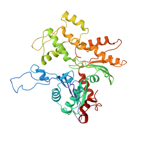
Near-atomic resolution for one state of f-actin.

Galkin, V.E.Orlova, A.Vos, M.R.Schroder, G.F.Egelman, E.H.

(2015) Structure 23: 173-182

  • DOI: https://doi.org/10.1016/j.str.2014.11.006
  • Primary Citation of Related Structures:  
    3J8I, 3J8J, 3J8K

  • PubMed Abstract: 

    Actin functions as a helical polymer, F-actin, but attempts to build an atomic model for this filament have been hampered by the fact that the filament cannot be crystallized and by structural heterogeneity. We have used a direct electron detector, cryo-electron microscopy, and the forces imposed on actin filaments in thin films to reconstruct one state of the filament at 4.7 Å resolution, which allows for building a reliable pseudo-atomic model of F-actin. We also report a different state of the filament where actin protomers adopt a conformation observed in the crystal structure of the G-actin-profilin complex with an open ATP-binding cleft. Comparison of the two structural states provides insights into ATP-hydrolysis and filament dynamics. The atomic model provides a framework for understanding why every buried residue in actin has been under intense selective pressure.


  • Organizational Affiliation

    Department of Physiological Sciences, Eastern Virginia Medical School, Norfolk, VA 23507, USA. Electronic address: galkinve@evms.edu.


Macromolecules
Find similar proteins by:  (by identity cutoff)  |  3D Structure
Entity ID: 1
MoleculeChains Sequence LengthOrganismDetailsImage
Actin, alpha skeletal muscleA [auth D],
B [auth E],
C [auth F],
D [auth G],
E [auth H]
377Oryctolagus cuniculusMutation(s): 0 
UniProt
Find proteins for P68135 (Oryctolagus cuniculus)
Explore P68135 
Go to UniProtKB:  P68135
Entity Groups  
Sequence Clusters30% Identity50% Identity70% Identity90% Identity95% Identity100% Identity
UniProt GroupP68135
Sequence Annotations
Expand
  • Reference Sequence
Small Molecules
Ligands 2 Unique
IDChains Name / Formula / InChI Key2D Diagram3D Interactions
ADP
Query on ADP

Download Ideal Coordinates CCD File 
F [auth D],
H [auth E],
J [auth F],
L [auth G],
N [auth H]
ADENOSINE-5'-DIPHOSPHATE
C10 H15 N5 O10 P2
XTWYTFMLZFPYCI-KQYNXXCUSA-N
MG
Query on MG

Download Ideal Coordinates CCD File 
G [auth D],
I [auth E],
K [auth F],
M [auth G],
O [auth H]
MAGNESIUM ION
Mg
JLVVSXFLKOJNIY-UHFFFAOYSA-N
Experimental Data & Validation

Experimental Data

  • Method: ELECTRON MICROSCOPY
  • Resolution: 4.70 Å
  • Aggregation State: FILAMENT 
  • Reconstruction Method: HELICAL 
EM Software:
TaskSoftware PackageVersion
RECONSTRUCTIONEMAN2
RECONSTRUCTIONSPIDER

Structure Validation

View Full Validation Report


Entry History 

Deposition Data

  • Released Date: 2015-01-14 
  • Deposition Author(s): Galkin, V.E.

Revision History  (Full details and data files)

  • Version 1.0: 2015-01-14
    Type: Initial release
  • Version 1.1: 2015-01-21
    Changes: Database references
  • Version 1.2: 2018-07-18
    Changes: Author supporting evidence, Data collection
  • Version 1.3: 2024-02-21
    Changes: Data collection, Database references, Derived calculations