Identification of the active sites in the methyltransferases of a transcribing dsRNA virus.
Zhu, B., Yang, C., Liu, H., Cheng, L., Song, F., Zeng, S., Huang, X., Ji, G., Zhu, P.(2014) J Mol Biology 426: 2167-2174
- PubMed: 24690366 Search on PubMedSearch on PubMed Central
- DOI: https://doi.org/10.1016/j.jmb.2014.03.013
- Primary Citation Related Structures:
3J6Q - PubMed Abstract:
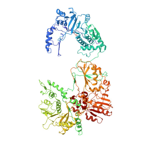
Many double-stranded RNA (dsRNA) viruses are capable of transcribing and capping RNA within a stable icosahedral viral capsid. The turret of turreted dsRNA viruses belonging to the family Reoviridae is formed by five copies of the turret protein, which contains domains with both 7-N-methyltransferase and 2'-O-methyltransferase activities, and serves to catalyze the methylation reactions during RNA capping. Cypovirus of the family Reoviridae provides a good model system for studying the methylation reactions in dsRNA viruses. Here, we present the structure of a transcribing cypovirus to a resolution of ~3.8Å by cryo-electron microscopy. The binding sites for both S-adenosyl-L-methionine and RNA in the two methyltransferases of the turret were identified. Structural analysis of the turret in complex with RNA revealed a pathway through which the RNA molecule reaches the active sites of the two methyltransferases before it is released into the cytoplasm. The pathway shows that RNA capping reactions occur in the active sites of different turret protein monomers, suggesting that RNA capping requires concerted efforts by at least three turret protein monomers. Thus, the turret structure provides novel insights into the precise mechanisms of RNA methylation.
- College of Physics and Information Science, Hunan Normal University, 36 Lushan Road, Changsha, Hunan 410081, China.
Organizational Affiliation: