3INK | pdb_00003ink

UNRAVELING THE STRUCTURE OF INTERLEUKIN-2: REPLY


Experimental Data Snapshot

  • Method: X-RAY DIFFRACTION
  • Resolution: 2.50 Å
  • R-Value Work: 
    0.202 (Depositor) 
  • R-Value Observed: 
    0.202 (Depositor) 

wwPDB Validation 3D Report Full Report

Validation slider image for 3INK

This is version 1.5 of the entry. See complete history

Literature

Response.

McKay, D.B.

(1992) Science 257: 412-413

Macromolecule Content 

  • Total Structure Weight: 30.81 kDa 
  • Atom Count: 1,996 
  • Modeled Residue Count: 244 
  • Deposited Residue Count: 266 
  • Unique protein chains: 1

Macromolecules

Find similar proteins by:|  3D Structure
Entity ID: 1
MoleculeChains  Sequence LengthOrganismDetailsImage
INTERLEUKIN-2A [auth C],
B [auth D]
133Homo sapiensMutation(s): 0 
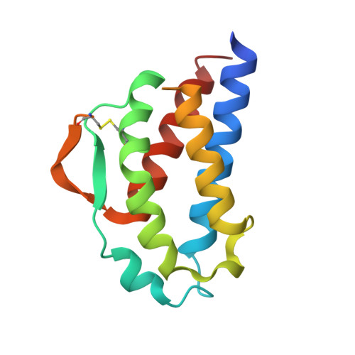
UniProt & NIH Common Fund Data Resources
Find proteins for P60568 (Homo sapiens)
Explore P60568 
Go to UniProtKB:  P60568
PHAROS:  P60568
GTEx:  ENSG00000109471 
Entity Groups
Sequence Clusters30% Identity50% Identity70% Identity90% Identity95% Identity100% Identity
UniProt GroupP60568
Sequence Annotations
Expand
Reference Sequence

Experimental Data & Validation

Experimental Data

  • Method: X-RAY DIFFRACTION
  • Resolution: 2.50 Å
  • R-Value Work:  0.202 (Depositor) 
  • R-Value Observed: 0.202 (Depositor) 
Space Group: P 1
Unit Cell:
Length ( Å )Angle ( ˚ )
a = 55.8α = 90
b = 40.1β = 109.3
c = 33.7γ = 93.2
Software Package:
Software NamePurpose
X-PLORmodel building
X-PLORrefinement
X-PLORphasing

Structure Validation

View Full Validation Report


Entry History 

Deposition Data

Revision History  (Full details and data files)

  • Version 1.0: 1993-10-31
    Type: Initial release
  • Version 1.1: 2008-03-25
    Changes: Version format compliance
  • Version 1.2: 2011-07-13
    Changes: Version format compliance
  • Version 1.3: 2013-09-04
    Changes: Database references
  • Version 1.4: 2024-06-05
    Changes: Data collection, Database references, Other
  • Version 1.5: 2024-10-30
    Changes: Structure summary