3IET | pdb_00003iet

Crystal Structure of 237mAb with antigen


Experimental Data Snapshot

  • Method: X-RAY DIFFRACTION
  • Resolution: 2.20 Å
  • R-Value Free: 
    0.271 (Depositor), 0.293 (DCC) 
  • R-Value Work: 
    0.227 (Depositor) 
  • R-Value Observed: 
    0.229 (Depositor) 

Starting Model: experimental
View more details

wwPDB Validation 3D Report Full Report

Validation slider image for 3IET

Ligand Structure Quality Assessment 


This is version 2.2 of the entry. See complete history

Literature

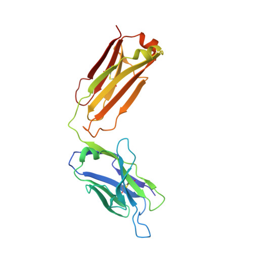
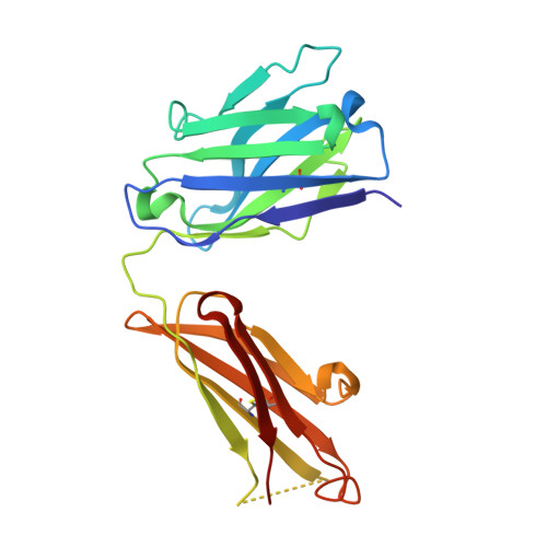
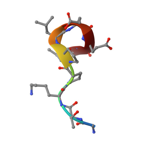
Antibody recognition of a unique tumor-specific glycopeptide antigen.

Brooks, C.L.Schietinger, A.Borisova, S.N.Kufer, P.Okon, M.Hirama, T.Mackenzie, C.R.Wang, L.X.Schreiber, H.Evans, S.V.

(2010) Proc Natl Acad Sci U S A 107: 10056-10061

  • DOI: https://doi.org/10.1073/pnas.0915176107
  • Primary Citation Related Structures: 
    3IET, 3IF1

  • PubMed Abstract: 

    Aberrant glycosylation and the overexpression of certain carbohydrate moieties is a consistent feature of cancers, and tumor-associated oligosaccharides are actively investigated as targets for immunotherapy. One of the most common aberrations in glycosylation patterns is the presentation of a single O-linked N-acetylgalactosamine on a threonine or serine residue known as the "Tn antigen." Whereas the ubiquitous nature of Tn antigens on cancers has made them a natural focus of vaccine research, such carbohydrate moieties are not always tumor-specific and have been observed on embryonic and nonmalignant adult tissue. Here we report the structural basis of binding of a complex of a monoclonal antibody (237mAb) with a truly tumor-specific glycopeptide containing the Tn antigen. In contrast to glycopeptide-specific antibodies in complex with simple peptides, 237mAb does not recognize a conformational epitope induced in the peptide by sugar substitution. Instead, 237mAb uses a pocket coded by germ-line genes to completely envelope the carbohydrate moiety itself while interacting with the peptide moiety in a shallow groove. Thus, 237mAb achieves its striking tumor specificity, with no observed physiological cross-reactivity to the unglycosylated peptide or the free glycan, by a combination of multiple weak but specific interactions to both the peptide and to the glycan portions of the antigen.


  • Organizational Affiliation
    • Department of Biochemistry and Microbiology, University of Victoria, Victoria, BC, Canada V8P 3P6.

Macromolecule Content 

  • Total Structure Weight: 97.4 kDa 
  • Atom Count: 7,204 
  • Modeled Residue Count: 868 
  • Deposited Residue Count: 888 
  • Unique protein chains: 3

Macromolecules

Find similar proteins by:|  3D Structure
Entity ID: 1
MoleculeChains  Sequence LengthOrganismDetailsImage
Immunoglobulin light chain (IgG2a)
A, C
217Mus musculusMutation(s): 0 
Find similar proteins by:|  3D Structure
Entity ID: 2
MoleculeChains  Sequence LengthOrganismDetailsImage
Immunoglobulin heavy chain (IgG2a)
B, D
218Mus musculusMutation(s): 0 
Find similar proteins by:  Sequence   |   3D Structure  
Entity ID: 3
MoleculeChains  Sequence LengthOrganismDetailsImage
PodoplaninE [auth X],
F [auth Q]
9Mus musculusMutation(s): 0 
Gene Names: PdpnRP23-348F1.2-002
UniProt & NIH Common Fund Data Resources
Find proteins for Q62011 (Mus musculus)
Explore Q62011 
Go to UniProtKB:  Q62011
IMPC:  MGI:103098
Entity Groups
UniProt GroupQ62011
Glycosylation
Glycosylation Sites: 1Go to GlyGen: Q62011-1
Sequence Annotations
Expand
Reference Sequence

Experimental Data & Validation

Experimental Data

  • Method: X-RAY DIFFRACTION
  • Resolution: 2.20 Å
  • R-Value Free:  0.271 (Depositor), 0.293 (DCC) 
  • R-Value Work:  0.227 (Depositor) 
  • R-Value Observed: 0.229 (Depositor) 
Space Group: C 1 2 1
Unit Cell:
Length ( Å )Angle ( ˚ )
a = 277.91α = 90
b = 38.24β = 108.99
c = 95.91γ = 90
Software Package:
Software NamePurpose
d*TREKdata processing
PHENIXrefinement
PDB_EXTRACTdata extraction
CrystalCleardata collection
d*TREKdata reduction
d*TREKdata scaling
PHASERphasing

Structure Validation

View Full Validation Report


Ligand Structure Quality Assessment 


Entry History 

Deposition Data

Revision History  (Full details and data files)

  • Version 1.0: 2010-06-23
    Type: Initial release
  • Version 1.1: 2011-07-13
    Changes: Version format compliance
  • Version 2.0: 2020-07-29
    Type: Remediation
    Reason: Carbohydrate remediation
    Changes: Atomic model, Data collection, Derived calculations, Structure summary
  • Version 2.1: 2023-09-06
    Changes: Data collection, Database references, Refinement description, Structure summary
  • Version 2.2: 2024-11-06
    Changes: Structure summary