3ICD

STRUCTURE OF A BACTERIAL ENZYME REGULATED BY PHOSPHORYLATION, ISOCITRATE DEHYDROGENASE


Experimental Data Snapshot

  • Method: X-RAY DIFFRACTION
  • Resolution: 2.50 Å
  • R-Value Work: 0.180 

wwPDB Validation   3D Report Full Report


This is version 1.4 of the entry. See complete history


Literature

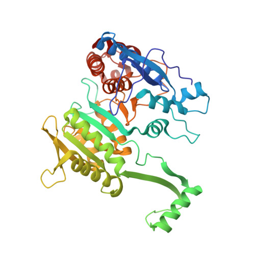
Structure of a bacterial enzyme regulated by phosphorylation, isocitrate dehydrogenase.

Hurley, J.H.Thorsness, P.E.Ramalingam, V.Helmers, N.H.Koshland Jr., D.E.Stroud, R.M.

(1989) Proc Natl Acad Sci U S A 86: 8635-8639

  • DOI: https://doi.org/10.1073/pnas.86.22.8635
  • Primary Citation of Related Structures:  
    3ICD

  • PubMed Abstract: 

    The structure of isocitrate dehydrogenase [threo-DS-isocitrate: NADP+ oxidoreductase (decarboxylating), EC 1.1.1.42] from Escherichia coli has been solved and refined at 2.5 A resolution and is topologically different from that of any other dehydrogenase. This enzyme, a dimer of identical 416-residue subunits, is inactivated by phosphorylation at Ser-113, which lies at the edge of an interdomain pocket that also contains many residues conserved between isocitrate dehydrogenase and isopropylmalate dehydrogenase. Isocitrate dehydrogenase contains an unusual clasp-like domain in which both polypeptide chains in the dimer interlock. Based on the structure of isocitrate dehydrogenase and conservation with isopropylmalate dehydrogenase, we suggest that the active site lies in an interdomain pocket close to the phosphorylation site.


  • Organizational Affiliation

    Department of Biochemistry and Biophysics, University of California, San Francisco 94143-0448.


Macromolecules
Find similar proteins by:  (by identity cutoff)  |  3D Structure
Entity ID: 1
MoleculeChains Sequence LengthOrganismDetailsImage
ISOCITRATE DEHYDROGENASE416Escherichia coliMutation(s): 0 
EC: 1.1.1.42
UniProt
Find proteins for P08200 (Escherichia coli (strain K12))
Explore P08200 
Go to UniProtKB:  P08200
Entity Groups  
Sequence Clusters30% Identity50% Identity70% Identity90% Identity95% Identity100% Identity
UniProt GroupP08200
Sequence Annotations
Expand
  • Reference Sequence
Experimental Data & Validation

Experimental Data

  • Method: X-RAY DIFFRACTION
  • Resolution: 2.50 Å
  • R-Value Work: 0.180 
  • Space Group: P 43 21 2
Unit Cell:
Length ( Å )Angle ( ˚ )
a = 105.1α = 90
b = 105.1β = 90
c = 150.3γ = 90
Software Package:
Software NamePurpose
X-PLORrefinement

Structure Validation

View Full Validation Report


Entry History 

Deposition Data

Revision History  (Full details and data files)

  • Version 1.0: 1991-01-15
    Type: Initial release
  • Version 1.1: 2008-03-25
    Changes: Version format compliance
  • Version 1.2: 2011-07-13
    Changes: Derived calculations, Version format compliance
  • Version 1.3: 2017-11-29
    Changes: Derived calculations, Other
  • Version 1.4: 2024-02-21
    Changes: Data collection, Database references