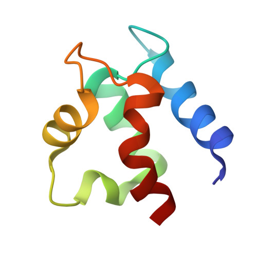
3ICB

THE REFINED STRUCTURE OF VITAMIN D-DEPENDENT CALCIUM-BINDING PROTEIN FROM BOVINE INTESTINE. MOLECULAR DETAILS, ION BINDING, AND IMPLICATIONS FOR THE STRUCTURE OF OTHER CALCIUM-BINDING PROTEINS


Experimental Data Snapshot

  • Method: X-RAY DIFFRACTION
  • Resolution: 2.30 Å
  • R-Value Work: 0.178 

wwPDB Validation   3D Report Full Report


This is version 1.2 of the entry. See complete history


Literature

The refined structure of vitamin D-dependent calcium-binding protein from bovine intestine. Molecular details, ion binding, and implications for the structure of other calcium-binding proteins.

Szebenyi, D.M.Moffat, K.

(1986) J Biol Chem 261: 8761-8777

  • Primary Citation of Related Structures:  
    3ICB

  • PubMed Abstract: 
  • The structure of bovine intestinal calcium-binding protein (ICaBP) has been determined crystallographically at a resolution of 2.3 A and refined by a least squares technique to an R factor of 17.8%. The refined structure includes all 600 non-hydrogen protein atoms, two bound calcium ions, and solvent consisting of one sulfate ion and 36 water molecules ...

    The structure of bovine intestinal calcium-binding protein (ICaBP) has been determined crystallographically at a resolution of 2.3 A and refined by a least squares technique to an R factor of 17.8%. The refined structure includes all 600 non-hydrogen protein atoms, two bound calcium ions, and solvent consisting of one sulfate ion and 36 water molecules. The molecule consists of two helix-loop-helix calcium-binding domains known as EF hands, connected by a linker containing a single turn of helix. Helix-helix interactions are primarily hydrophobic, but also include a few strategic hydrogen bonds. Most of the hydrogen bonds, however, are found in the calcium-binding loops, where they occur both within a single loop and between the two. Examination of the hydrogen bonding patterns in the calcium-binding loops of ICaBP and the related protein, parvalbumin, reveals several conserved hydrogen bonds which are evidently important for loop stabilization. The primary and tertiary structural features which promote the formation of an EF hand were originally identified from the structure of parvalbumin. They are modified in light of the ICaBP structure and considered as they apply to other calcium-binding proteins. The C-terminal domain of ICaBP is a normal EF hand, with ion binding properties similar to those of the calmodulin hands, but the N-terminal domain is a variant hand whose calcium ligands are mostly peptide carbonyls. Relative to a normal EF hand, this domain exhibits a similar KD for calcium binding but a greatly reduced affinity for calcium analogs such as cadmium and the lanthanide series. Lanthanides in particular may be inappropriate models for calcium in this system.


    Related Citations: 
    • Structure of Vitamin D-Dependent Calcium-Binding Protein from Bovine Intestine
      Szebenyi, D.M.E., Obendorf, S.K., Moffat, K.
      (1981) Nature 294: 327
    • Preliminary Crystallographic Data for a Calcium Binding Protein from Bovine Intestine
      Moffat, K., Fullmer, C.H., Wasserman, R.H.
      (1975) J Mol Biol 97: 661
    • The Amino Acid Sequence of Bovine Intestinal Calcium-Binding Protein
      Fullmer, C.S., Wasserman, R.H.
      (1981) J Biol Chem 256: 5669

    Organizational Affiliation

    Kristallographie, Institut für Chemie und Biochemie, Freie Universität Berlin, Takustrasse 6, Berlin 14195, Germany.


Macromolecules
Find similar proteins by:  (by identity cutoff)  |  3D Structure
Entity ID: 1
MoleculeChainsSequence LengthOrganismDetailsImage
CALCIUM-BINDING PROTEINA75Bos taurusMutation(s): 0 
Gene Names: S100GCALB3S100D
UniProt
Find proteins for P02633 (Bos taurus)
Explore P02633 
Go to UniProtKB:  P02633
Entity Groups  
Sequence Clusters30% Identity50% Identity70% Identity90% Identity95% Identity100% Identity
UniProt GroupP02633
Protein Feature View
Expand
  • Reference Sequence
Small Molecules
Ligands 2 Unique
IDChainsName / Formula / InChI Key2D Diagram3D Interactions
SO4
Query on SO4

Download Ideal Coordinates CCD File 
D [auth A]SULFATE ION
O4 S
QAOWNCQODCNURD-UHFFFAOYSA-L
 Ligand Interaction
CA
Query on CA

Download Ideal Coordinates CCD File 
B [auth A],
C [auth A]
CALCIUM ION
Ca
BHPQYMZQTOCNFJ-UHFFFAOYSA-N
 Ligand Interaction
Experimental Data & Validation

Experimental Data

  • Method: X-RAY DIFFRACTION
  • Resolution: 2.30 Å
  • R-Value Work: 0.178 
  • Space Group: P 21 21 2
Unit Cell:
Length ( Å )Angle ( ˚ )
a = 56.2α = 90
b = 42.6β = 90
c = 29.2γ = 90
Software Package:
Software NamePurpose
PROLSQrefinement

Structure Validation

View Full Validation Report


Entry History 

Deposition Data

Revision History  (Full details and data files)

  • Version 1.0: 1986-10-24
    Type: Initial release
  • Version 1.1: 2008-03-03
    Changes: Version format compliance
  • Version 1.2: 2011-07-13
    Changes: Version format compliance