3IA3

A cis-proline in alpha-hemoglobin stabilizing Protein directs the structural reorganization of alpha-hemoglobin


Experimental Data Snapshot

  • Method: X-RAY DIFFRACTION
  • Resolution: 3.20 Å
  • R-Value Free: 0.293 
  • R-Value Work: 0.230 
  • R-Value Observed: 0.230 

wwPDB Validation   3D Report Full Report


Ligand Structure Quality Assessment 


This is version 1.2 of the entry. See complete history


Literature

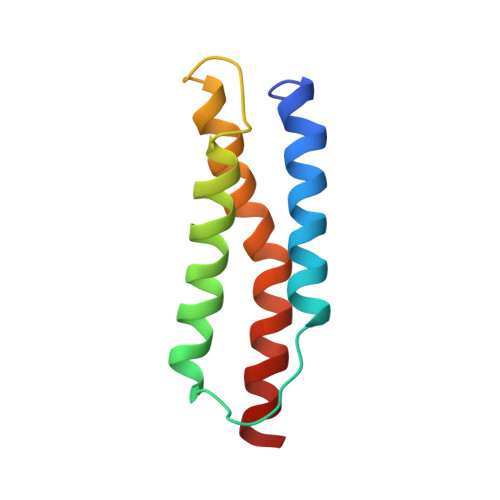
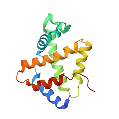
A cis-proline in alpha-hemoglobin stabilizing protein directs the structural reorganization of alpha-hemoglobin.

Gell, D.A.Feng, L.Zhou, S.Jeffrey, P.D.Bendak, K.Gow, A.Weiss, M.J.Shi, Y.Mackay, J.P.

(2009) J Biol Chem 284: 29462-29469

  • DOI: https://doi.org/10.1074/jbc.M109.027045
  • Primary Citation of Related Structures:  
    3IA3

  • PubMed Abstract: 

    alpha-Hemoglobin (alphaHb) stabilizing protein (AHSP) is expressed in erythropoietic tissues as an accessory factor in hemoglobin synthesis. AHSP forms a specific complex with alphaHb and suppresses the heme-catalyzed evolution of reactive oxygen species by converting alphaHb to a conformation in which the heme is coordinated at both axial positions by histidine side chains (bis-histidyl coordination). Currently, the detailed mechanism by which AHSP induces structural changes in alphaHb has not been determined. Here, we present x-ray crystallography, NMR spectroscopy, and mutagenesis data that identify, for the first time, the importance of an evolutionarily conserved proline, Pro(30), in loop 1 of AHSP. Mutation of Pro(30) to a variety of residue types results in reduced ability to convert alphaHb. In complex with alphaHb, AHSP Pro(30) adopts a cis-peptidyl conformation and makes contact with the N terminus of helix G in alphaHb. Mutations that stabilize the cis-peptidyl conformation of free AHSP, also enhance the alphaHb conversion activity. These findings suggest that AHSP loop 1 can transmit structural changes to the heme pocket of alphaHb, and, more generally, highlight the importance of cis-peptidyl prolyl residues in defining the conformation of regulatory protein loops.


  • Organizational Affiliation

    School of Molecular and Microbial Biosciences, University of Sydney, New South Wales 2006, Australia. dagell@mail.usyd.edu.au


Macromolecules
Find similar proteins by:  (by identity cutoff)  |  3D Structure
Entity ID: 1
MoleculeChains Sequence LengthOrganismDetailsImage
Alpha-hemoglobin-stabilizing protein
A, C
91Homo sapiensMutation(s): 0 
Gene Names: AHSPEDRFERAF
UniProt & NIH Common Fund Data Resources
Find proteins for Q9NZD4 (Homo sapiens)
Explore Q9NZD4 
Go to UniProtKB:  Q9NZD4
PHAROS:  Q9NZD4
GTEx:  ENSG00000169877 
Entity Groups  
Sequence Clusters30% Identity50% Identity70% Identity90% Identity95% Identity100% Identity
UniProt GroupQ9NZD4
Sequence Annotations
Expand
  • Reference Sequence
Find similar proteins by:  (by identity cutoff)  |  3D Structure
Entity ID: 2
MoleculeChains Sequence LengthOrganismDetailsImage
Hemoglobin subunit alpha
B, D
145Homo sapiensMutation(s): 0 
Gene Names: HBA1HBA2
UniProt & NIH Common Fund Data Resources
Find proteins for P69905 (Homo sapiens)
Explore P69905 
Go to UniProtKB:  P69905
PHAROS:  P69905
Entity Groups  
Sequence Clusters30% Identity50% Identity70% Identity90% Identity95% Identity100% Identity
UniProt GroupP69905
Sequence Annotations
Expand
  • Reference Sequence
Small Molecules
Ligands 1 Unique
IDChains Name / Formula / InChI Key2D Diagram3D Interactions
HEM
Query on HEM

Download Ideal Coordinates CCD File 
E [auth B],
F [auth D]
PROTOPORPHYRIN IX CONTAINING FE
C34 H32 Fe N4 O4
KABFMIBPWCXCRK-RGGAHWMASA-L
Experimental Data & Validation

Experimental Data

  • Method: X-RAY DIFFRACTION
  • Resolution: 3.20 Å
  • R-Value Free: 0.293 
  • R-Value Work: 0.230 
  • R-Value Observed: 0.230 
  • Space Group: P 61 2 2
Unit Cell:
Length ( Å )Angle ( ˚ )
a = 65.23α = 90
b = 65.23β = 90
c = 439.66γ = 120
Software Package:
Software NamePurpose
CBASSdata collection
PHASERphasing
CNSrefinement
DENZOdata reduction
SCALEPACKdata scaling

Structure Validation

View Full Validation Report


Ligand Structure Quality Assessment 


Entry History 

Deposition Data

Revision History  (Full details and data files)

  • Version 1.0: 2009-07-28
    Type: Initial release
  • Version 1.1: 2011-07-13
    Changes: Version format compliance
  • Version 1.2: 2024-02-21
    Changes: Data collection, Database references, Derived calculations