3I6Z | pdb_00003i6z

3D Structure of Torpedo californica acetylcholinesterase complexed with N-saccharinohexyl-galanthamine


Experimental Data Snapshot

  • Method: X-RAY DIFFRACTION
  • Resolution: 2.19 Å
  • R-Value Free: 
    0.212 (Depositor), 0.214 (DCC) 
  • R-Value Work: 
    0.183 (Depositor), 0.187 (DCC) 
  • R-Value Observed: 
    0.183 (Depositor) 

Starting Model: experimental
View more details

wwPDB Validation 3D Report Full Report

Validation slider image for 3I6Z

Ligand Structure Quality Assessment 


This is version 2.2 of the entry. See complete history

Literature

Probing Torpedo californica acetylcholinesterase catalytic gorge with two novel bis-functional galanthamine derivatives.

Bartolucci, C.Haller, L.A.Jordis, U.Fels, G.Lamba, D.

(2010) J Med Chem 53: 745-751

  • DOI: https://doi.org/10.1021/jm901296p
  • Primary Citation Related Structures: 
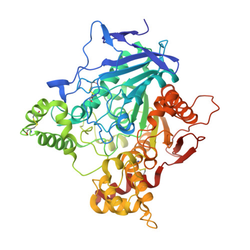
    3I6M, 3I6Z

  • PubMed Abstract: 

    N-Piperidinopropyl-galanthamine (2) and N-saccharinohexyl-galanthamine (3) were used to investigate interaction sites along the active site gorge of Torpedo californica actylcholinesterase (TcAChE). The crystal structure of TcAChE-2 solved at 2.3 A showed that the N-piperidinopropyl group in 2 is not stretched along the gorge but is folded over the galanthamine moiety. This result was unexpected because the three carbon alkyl chain is just long enough for the bulky piperidine group to be placed above the bottleneck (Tyr121, Phe330) midway down the gorge. The crystal structure of TcAChE-3 at 2.2 A confirmed that a dual interaction with the sites at the bottom, and at the entrance of the gorge, enhances inhibitory activity: a chain of six carbon atoms has, in this class of derivatives, the correct length for optimal interactions with the peripheral anionic site (PAS).


  • Organizational Affiliation
    • Istituto di Cristallografia, Consiglio Nazionale delle Ricerche, Area della Ricerca di Roma, P.O. Box 10, I-00016 Monterotondo Stazione(Roma), Italy.

Macromolecule Content 

  • Total Structure Weight: 61.63 kDa 
  • Atom Count: 4,699 
  • Modeled Residue Count: 532 
  • Deposited Residue Count: 534 
  • Unique protein chains: 1

Macromolecules

Find similar proteins by:|  3D Structure
Entity ID: 1
MoleculeChains  Sequence LengthOrganismDetailsImage
Acetylcholinesterase534Tetronarce californicaMutation(s): 0 
EC: 3.1.1.7
UniProt
Find proteins for P04058 (Tetronarce californica)
Explore P04058 
Go to UniProtKB:  P04058
Entity Groups
Sequence Clusters30% Identity50% Identity70% Identity90% Identity95% Identity100% Identity
UniProt GroupP04058
Glycosylation
Glycosylation Sites: 2
Sequence Annotations
Expand
Reference Sequence

Oligosaccharides

Help  
Entity ID: 2
MoleculeChains Length2D Diagram GlycosylationD Interactions
2-acetamido-2-deoxy-beta-D-glucopyranose-(1-4)-2-acetamido-2-deoxy-beta-D-glucopyranose
B
2N-Glycosylation
Glycosylation Resources
GlyTouCan: G42666HT
GlyCosmos: G42666HT
GlyGen: G42666HT

Small Molecules

Ligands 2 Unique
IDChains Name / Formula / InChI Key2D Diagram3D Interactions
G6X

Query on G6X


Download:Ideal Coordinates CCD File
D [auth A]2-{6-[(4aS,6R,8aS)-6-hydroxy-3-methoxy-5,6,9,10-tetrahydro-4aH-[1]benzofuro[3a,3,2-ef][2]benzazepin-11(12H)-yl]hexyl}-1 ,2-benzisothiazol-3(2H)-one 1,1-dioxide
C29 H34 N2 O6 S
VAEMEXGGRSPJEF-LOMAHXHCSA-N
NAG

Query on NAG


Download:Ideal Coordinates CCD File
C [auth A]2-acetamido-2-deoxy-beta-D-glucopyranose
C8 H15 N O6
OVRNDRQMDRJTHS-FMDGEEDCSA-N

Experimental Data & Validation

Experimental Data

  • Method: X-RAY DIFFRACTION
  • Resolution: 2.19 Å
  • R-Value Free:  0.212 (Depositor), 0.214 (DCC) 
  • R-Value Work:  0.183 (Depositor), 0.187 (DCC) 
  • R-Value Observed: 0.183 (Depositor) 
Space Group: P 31 2 1
Unit Cell:
Length ( Å )Angle ( ˚ )
a = 111.339α = 90
b = 111.339β = 90
c = 137.207γ = 120
Software Package:
Software NamePurpose
MAR345data collection
AMoREphasing
CNSrefinement
DENZOdata reduction
SCALEPACKdata scaling

Structure Validation

View Full Validation Report


Ligand Structure Quality Assessment 


Entry History 

Deposition Data

Revision History  (Full details and data files)

  • Version 1.0: 2010-01-12
    Type: Initial release
  • Version 1.1: 2011-07-13
    Changes: Version format compliance
  • Version 1.2: 2017-11-01
    Changes: Refinement description
  • Version 2.0: 2020-07-29
    Type: Remediation
    Reason: Carbohydrate remediation
    Changes: Atomic model, Data collection, Derived calculations, Structure summary
  • Version 2.1: 2023-09-06
    Changes: Data collection, Database references, Refinement description, Structure summary
  • Version 2.2: 2024-11-06
    Changes: Structure summary