3I17 | pdb_00003i17

Crystal structure of the apo R132K:L121E mutant of cellular retinoic acid-binding protein II at 1.68 angstrom resolution


Experimental Data Snapshot

  • Method: X-RAY DIFFRACTION
  • Resolution: 1.68 Å
  • R-Value Free: 
    0.231 (Depositor), 0.242 (DCC) 
  • R-Value Work: 
    0.180 (Depositor) 
  • R-Value Observed: 
    0.183 (Depositor) 

Starting Model: experimental
View more details

wwPDB Validation 3D Report Full Report

Validation slider image for 3I17

This is version 1.4 of the entry. See complete history

Literature

Elucidating the exact role of engineered CRABPII residues for the formation of a retinal protonated Schiff base.

Vasileiou, C.Wang, W.Jia, X.Lee, K.S.Watson, C.T.Geiger, J.H.Borhan, B.

(2009) Proteins 77: 812-822

  • DOI: https://doi.org/10.1002/prot.22495
  • Primary Citation Related Structures: 
    3I17

  • PubMed Abstract: 

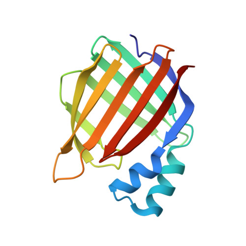
    Cellular Retinoic Acid Binding Protein II (CRABPII) has been reengineered to specifically bind and react with all-trans-retinal to form a protonated Schiff base. Each step of this process has been dissected and four residues (Lys132, Tyr134, Arg111, and Glu121) within the CRABPII binding site have been identified as crucial for imine formation and/or protonation. The precise role of each residue has been examined through site directed mutagenesis and crystallographic studies. The crystal structure of the R132K:L121E-CRABPII (PDB-3I17) double mutant suggests a direct interaction between engineered Glu121 and the native Arg111, which is critical for both Schiff base formation and protonation.


  • Organizational Affiliation
    • Department of Chemistry, Michigan State University, East Lansing, Michigan 48824, USA.

Macromolecule Content 

  • Total Structure Weight: 31.14 kDa 
  • Atom Count: 2,455 
  • Modeled Residue Count: 274 
  • Deposited Residue Count: 274 
  • Unique protein chains: 1

Macromolecules

Find similar proteins by:|  3D Structure
Entity ID: 1
MoleculeChains  Sequence LengthOrganismDetailsImage
Cellular retinoic acid-binding protein 2A [auth B],
B [auth A]
137Homo sapiensMutation(s): 2 
Gene Names: CRABP2
UniProt & NIH Common Fund Data Resources
Find proteins for P29373 (Homo sapiens)
Explore P29373 
Go to UniProtKB:  P29373
PHAROS:  P29373
GTEx:  ENSG00000143320 
Entity Groups
Sequence Clusters30% Identity50% Identity70% Identity90% Identity95% Identity100% Identity
UniProt GroupP29373
Sequence Annotations
Expand
Reference Sequence

Experimental Data & Validation

Experimental Data

  • Method: X-RAY DIFFRACTION
  • Resolution: 1.68 Å
  • R-Value Free:  0.231 (Depositor), 0.242 (DCC) 
  • R-Value Work:  0.180 (Depositor) 
  • R-Value Observed: 0.183 (Depositor) 
Space Group: P 1
Unit Cell:
Length ( Å )Angle ( ˚ )
a = 34.699α = 105.8
b = 37.271β = 106.44
c = 60.371γ = 89.97
Software Package:
Software NamePurpose
HKL-2000data collection
MOLREPphasing
REFMACrefinement
HKL-2000data reduction
HKL-2000data scaling

Structure Validation

View Full Validation Report


Entry History 

Deposition Data

Revision History  (Full details and data files)

  • Version 1.0: 2009-07-28
    Type: Initial release
  • Version 1.1: 2011-07-13
    Changes: Version format compliance
  • Version 1.2: 2016-12-28
    Changes: Structure summary
  • Version 1.3: 2021-10-13
    Changes: Database references
  • Version 1.4: 2023-09-06
    Changes: Data collection, Refinement description