3I0W | pdb_00003i0w

Crystal structure of Clostridium acetobutylicum 8-oxoguanine glycosylase/lyase in complex with dsDNA containing cytosine opposite to 8-oxoG


Experimental Data Snapshot

  • Method: X-RAY DIFFRACTION
  • Resolution: 1.73 Å
  • R-Value Free: 
    0.219 (Depositor), 0.224 (DCC) 
  • R-Value Work: 
    0.195 (Depositor), 0.203 (DCC) 
  • R-Value Observed: 
    0.195 (Depositor) 

Starting Model: experimental
View more details

wwPDB Validation 3D Report Full Report

Validation slider image for 3I0W

This is version 1.3 of the entry. See complete history

Literature

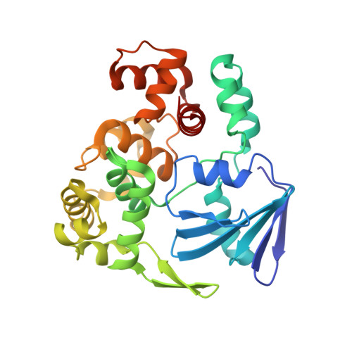
Structural basis for the lack of opposite base specificity of Clostridium acetobutylicum 8-oxoguanine DNA glycosylase.

Faucher, F.Wallace, S.S.Doublie, S.

(2009) DNA Repair (Amst) 8: 1283-1289

  • DOI: https://doi.org/10.1016/j.dnarep.2009.08.002
  • Primary Citation Related Structures: 
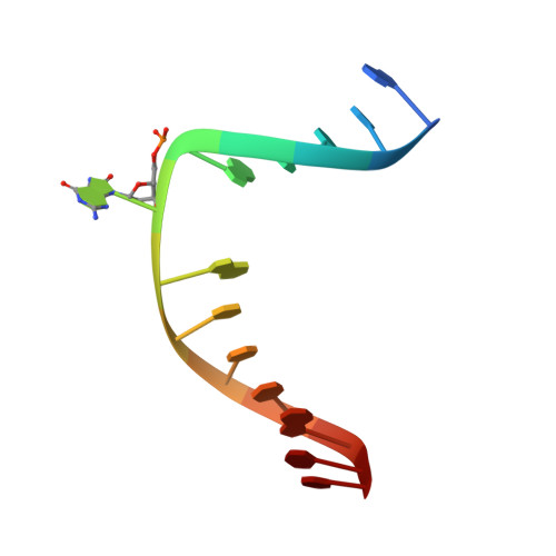
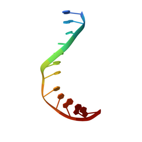
    3I0W, 3I0X

  • PubMed Abstract: 

    7,8-Dihydro-8-oxoguanine (8-oxoG) is the major oxidative product of guanine and the most prevalent base lesion observed in DNA molecules. Because 8-oxoG has the capability to form a Hoogsteen pair with adenine (8-oxoG:A) in addition to a normal Watson-Crick pair with cytosine (8-oxoG:C), this lesion can lead to a G:C-->T:A transversion after replication. However, 8-oxoG is recognized and excised by the 8-oxoguanine DNA glycosylase (Ogg) of the base excision repair pathway. Members of the Ogg1 family usually display a strong preference for a C opposite the lesion. In contrast, the atypical Ogg1 from Clostridium actetobutylicum (CacOgg) can excise 8-oxoG when paired with either one of the four bases, albeit with a preference for C and A. Here we describe the first high-resolution crystal structures of CacOgg in complex with duplex DNA containing the 8-oxoG lesion paired to cytosine and to adenine. A structural comparison with human OGG1 provides a rationale for the lack of opposite base specificity displayed by the bacterial Ogg.


  • Organizational Affiliation
    • Department of Microbiology and Molecular Genetics, The Markey Center for Molecular Genetics, University of Vermont, Stafford Hall, 95 Carrigan Drive, Burlington, VT 05405-0068, USA.

Macromolecule Content 

  • Total Structure Weight: 42.21 kDa 
  • Atom Count: 3,302 
  • Modeled Residue Count: 315 
  • Deposited Residue Count: 315 
  • Unique protein chains: 1
  • Unique nucleic acid chains: 2

Macromolecules


Find similar proteins by:|  3D Structure
Entity ID: 1
MoleculeChains  Sequence LengthOrganismDetailsImage
8-oxoguanine-DNA-glycosylase290Clostridium acetobutylicumMutation(s): 1 
Gene Names: CAC2707CA_C2707
EC: 3.2.2 (PDB Primary Data), 4.2.99.18 (PDB Primary Data)
UniProt
Find proteins for Q97FM4 (Clostridium acetobutylicum (strain ATCC 824 / DSM 792 / JCM 1419 / IAM 19013 / LMG 5710 / NBRC 13948 / NRRL B-527 / VKM B-1787 / 2291 / W))
Explore Q97FM4 
Go to UniProtKB:  Q97FM4
Entity Groups
Sequence Clusters30% Identity50% Identity70% Identity90% Identity95% Identity100% Identity
UniProt GroupQ97FM4
Sequence Annotations
Expand
Reference Sequence
Find similar nucleic acids by:  Sequence
Entity ID: 2
MoleculeChains LengthOrganismImage
5'-D(*AP*TP*CP*CP*AP*(8OG)P*GP*TP*CP*TP*AP*CP*C)-3'13N/A
Sequence Annotations
Expand
Reference Sequence
Find similar nucleic acids by:  Sequence
Entity ID: 3
MoleculeChains LengthOrganismImage
5'-D(*GP*GP*TP*AP*GP*AP*CP*CP*TP*GP*GP*A)-3'12N/A
Sequence Annotations
Expand
Reference Sequence

Experimental Data & Validation

Experimental Data

  • Method: X-RAY DIFFRACTION
  • Resolution: 1.73 Å
  • R-Value Free:  0.219 (Depositor), 0.224 (DCC) 
  • R-Value Work:  0.195 (Depositor), 0.203 (DCC) 
  • R-Value Observed: 0.195 (Depositor) 
Space Group: P 65 2 2
Unit Cell:
Length ( Å )Angle ( ˚ )
a = 92.4α = 90
b = 92.4β = 90
c = 191.05γ = 120
Software Package:
Software NamePurpose
XSCALEdata scaling
MOLREPphasing
CNSrefinement
PDB_EXTRACTdata extraction
MAR345dtbdata collection
XDSdata reduction

Structure Validation

View Full Validation Report


Entry History 

Deposition Data

Revision History  (Full details and data files)

  • Version 1.0: 2009-09-29
    Type: Initial release
  • Version 1.1: 2011-07-13
    Changes: Version format compliance
  • Version 1.2: 2021-10-13
    Changes: Database references, Derived calculations
  • Version 1.3: 2023-09-06
    Changes: Data collection, Refinement description