3I06

Crystal structure of cruzain covalently bound to a purine nitrile


Experimental Data Snapshot

  • Method: X-RAY DIFFRACTION
  • Resolution: 1.10 Å
  • R-Value Free: 0.142 
  • R-Value Work: 0.119 
  • R-Value Observed: 0.120 

wwPDB Validation   3D Report Full Report


Ligand Structure Quality Assessment 


This is version 1.2 of the entry. See complete history


Literature

Identification and optimization of inhibitors of trypanosomal cysteine proteases: cruzain, rhodesain, and TbCatB.

Mott, B.T.Ferreira, R.S.Simeonov, A.Jadhav, A.Ang, K.K.Leister, W.Shen, M.Silveira, J.T.Doyle, P.S.Arkin, M.R.McKerrow, J.H.Inglese, J.Austin, C.P.Thomas, C.J.Shoichet, B.K.Maloney, D.J.

(2010) J Med Chem 53: 52-60

  • DOI: https://doi.org/10.1021/jm901069a
  • Primary Citation of Related Structures:  
    3I06

  • PubMed Abstract: 

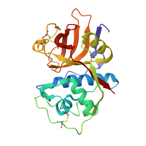
    Trypanosoma cruzi and Trypanosoma brucei are parasites that cause Chagas' disease and African sleeping sickness, respectively. Both parasites rely on essential cysteine proteases for survival: cruzain for T. cruzi and TbCatB/rhodesain for T. brucei. A recent quantitative high-throughput screen of cruzain identified triazine nitriles, which are known inhibitors of other cysteine proteases, as reversible inhibitors of the enzyme. Structural modifications detailed herein, including core scaffold modification from triazine to purine, improved the in vitro potency against both cruzain and rhodesain by 350-fold, while also gaining activity against T. brucei parasites. Selected compounds were screened against a panel of human cysteine and serine proteases to determine selectivity, and a cocrystal was obtained of our most potent analogue bound to cruzain.


  • Organizational Affiliation

    NIH Chemical Genomics Center, National Human Genome Research Institute, National Institutes of Health, 9800 Medical Center Drive, MSC 3370 Bethesda, Maryland 20892-3370, USA.


Macromolecules
Find similar proteins by:  (by identity cutoff)  |  3D Structure
Entity ID: 1
MoleculeChains Sequence LengthOrganismDetailsImage
Cruzipain215Trypanosoma cruziMutation(s): 0 
EC: 3.4.22.51
UniProt
Find proteins for P25779 (Trypanosoma cruzi)
Explore P25779 
Go to UniProtKB:  P25779
Entity Groups  
Sequence Clusters30% Identity50% Identity70% Identity90% Identity95% Identity100% Identity
UniProt GroupP25779
Sequence Annotations
Expand
  • Reference Sequence
Small Molecules
Ligands 1 Unique
IDChains Name / Formula / InChI Key2D Diagram3D Interactions
QL2
Query on QL2

Download Ideal Coordinates CCD File 
B [auth A]6-[(3,5-difluorophenyl)amino]-9-ethyl-9H-purine-2-carbonitrile
C14 H10 F2 N6
SZYYBVWPURUFRR-UHFFFAOYSA-N
Binding Affinity Annotations 
IDSourceBinding Affinity
QL2 PDBBind:  3I06 IC50: 0.2 (nM) from 1 assay(s)
BindingDB:  3I06 IC50: min: 0.2, max: 10 (nM) from 2 assay(s)
Experimental Data & Validation

Experimental Data

  • Method: X-RAY DIFFRACTION
  • Resolution: 1.10 Å
  • R-Value Free: 0.142 
  • R-Value Work: 0.119 
  • R-Value Observed: 0.120 
  • Space Group: P 65 2 2
Unit Cell:
Length ( Å )Angle ( ˚ )
a = 82.967α = 90
b = 82.967β = 90
c = 101.895γ = 120
Software Package:
Software NamePurpose
MOSFLMdata reduction
SCALAdata scaling
PHASERphasing
REFMACrefinement
PDB_EXTRACTdata extraction

Structure Validation

View Full Validation Report


Ligand Structure Quality Assessment 


Entry History 

Deposition Data

Revision History  (Full details and data files)

  • Version 1.0: 2009-12-15
    Type: Initial release
  • Version 1.1: 2011-07-13
    Changes: Version format compliance
  • Version 1.2: 2023-09-06
    Changes: Data collection, Database references, Derived calculations, Refinement description