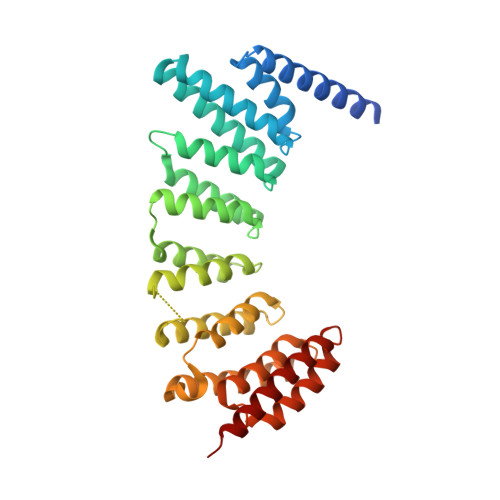
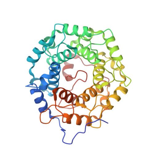
3HXC

Engineered RabGGTase in complex with a peptidomimetic inhibitor (compound 8)


Experimental Data Snapshot

  • Method: X-RAY DIFFRACTION
  • Resolution: 1.95 Å
  • R-Value Free: 0.218 
  • R-Value Work: 0.169 
  • R-Value Observed: 0.172 

wwPDB Validation   3D Report Full Report


Ligand Structure Quality Assessment 


This is version 1.4 of the entry. See complete history


Literature

Design, synthesis, and characterization of Peptide-based rab geranylgeranyl transferase inhibitors

Tan, K.T.Guiu-Rozas, E.Bon, R.S.Guo, Z.Delon, C.Wetzel, S.Arndt, S.Alexandrov, K.Waldmann, H.Goody, R.S.Wu, Y.W.Blankenfeldt, W.

(2009) J Med Chem 52: 8025-8037

  • DOI: https://doi.org/10.1021/jm901117d
  • Primary Citation of Related Structures:  
    3HXB, 3HXC, 3HXD, 3HXE, 3HXF

  • PubMed Abstract: 

    Rab geranylgeranyl transferase (RabGGTase) catalyzes the attachment of geranylgeranyl isoprenoids to Rab guanine triphosphatases, which are key regulators in vesicular transport. Because geranylgeranylation is required for proper function and overexpression of Rabs has been observed in various cancers, RabGGTase may be a target for novel therapeutics. The development of selective inhibitors is, however, difficult because two related enzymes involved in other cellular processes exist in eukaryotes and because RabGGTase recognizes protein substrates indirectly, resulting in relaxed specificity. We report the synthesis of a peptidic library based on the farnesyl transferase inhibitor pepticinnamin E. Of 469 compounds investigated, several were identified as selective for RabGGTase with low micromolar IC(50) values. The compounds were not generally cytotoxic and inhibited Rab isoprenylation in COS-7 cells. Crystal structure analysis revealed that selective inhibitors interact with a tunnel unique to RabGGTase, implying that this structural motif is an attractive target for improved RabGGTase inhibitors.


  • Organizational Affiliation

    Department of Chemical Biology, Biochemistry, Max Planck Institute of Molecular Physiology,Otto-Hahn-Strassse 11, 44227 Dortmund, Germany.


Macromolecules
Find similar proteins by:  (by identity cutoff)  |  3D Structure
Entity ID: 1
MoleculeChains Sequence LengthOrganismDetailsImage
Geranylgeranyl transferase type-2 subunit alpha331Rattus norvegicusMutation(s): 0 
EC: 2.5.1.60
UniProt
Find proteins for Q08602 (Rattus norvegicus)
Explore Q08602 
Go to UniProtKB:  Q08602
Entity Groups  
Sequence Clusters30% Identity50% Identity70% Identity90% Identity95% Identity100% Identity
UniProt GroupQ08602
Sequence Annotations
Expand
  • Reference Sequence
Find similar proteins by:  (by identity cutoff)  |  3D Structure
Entity ID: 2
MoleculeChains Sequence LengthOrganismDetailsImage
Geranylgeranyl transferase type-2 subunit beta331Rattus norvegicusMutation(s): 0 
EC: 2.5.1.60
UniProt
Find proteins for Q08603 (Rattus norvegicus)
Explore Q08603 
Go to UniProtKB:  Q08603
Entity Groups  
Sequence Clusters30% Identity50% Identity70% Identity90% Identity95% Identity100% Identity
UniProt GroupQ08603
Sequence Annotations
Expand
  • Reference Sequence
Small Molecules
Ligands 3 Unique
IDChains Name / Formula / InChI Key2D Diagram3D Interactions
BD6
Query on BD6

Download Ideal Coordinates CCD File 
E [auth B]N-[(benzyloxy)carbonyl]-L-histidyl-N-methyl-L-phenylalanyl-N-hydroxy-L-tryptophanamide
C35 H37 N7 O6
PUYLOEIRYPDFJO-CHQNGUEUSA-N
ZN
Query on ZN

Download Ideal Coordinates CCD File 
C [auth B]ZINC ION
Zn
PTFCDOFLOPIGGS-UHFFFAOYSA-N
CA
Query on CA

Download Ideal Coordinates CCD File 
D [auth B]CALCIUM ION
Ca
BHPQYMZQTOCNFJ-UHFFFAOYSA-N
Binding Affinity Annotations 
IDSourceBinding Affinity
BD6 PDBBind:  3HXC IC50: 5200 (nM) from 1 assay(s)
Experimental Data & Validation

Experimental Data

  • Method: X-RAY DIFFRACTION
  • Resolution: 1.95 Å
  • R-Value Free: 0.218 
  • R-Value Work: 0.169 
  • R-Value Observed: 0.172 
  • Space Group: P 21 21 21
Unit Cell:
Length ( Å )Angle ( ˚ )
a = 66.775α = 90
b = 90.616β = 90
c = 114.699γ = 90
Software Package:
Software NamePurpose
PHASERphasing
REFMACrefinement
XDSdata reduction
XSCALEdata scaling

Structure Validation

View Full Validation Report


Ligand Structure Quality Assessment 


Entry History 

Deposition Data

Revision History  (Full details and data files)

  • Version 1.0: 2009-09-08
    Type: Initial release
  • Version 1.1: 2011-07-13
    Changes: Version format compliance
  • Version 1.2: 2013-02-13
    Changes: Refinement description
  • Version 1.3: 2017-07-26
    Changes: Refinement description, Source and taxonomy
  • Version 1.4: 2024-02-21
    Changes: Data collection, Database references, Derived calculations