3HSP

Ternary structure of neuronal nitric oxide synthase with NHA and NO bound(2)


Experimental Data Snapshot

  • Method: X-RAY DIFFRACTION
  • Resolution: 2.20 Å
  • R-Value Free: 0.217 
  • R-Value Work: 0.164 
  • R-Value Observed: 0.167 

wwPDB Validation   3D Report Full Report


Ligand Structure Quality Assessment 


This is version 1.3 of the entry. See complete history


Literature

Single crystal structural and absorption spectral characterizations of nitric oxide synthase complexed with N(omega)-hydroxy-L-arginine and diatomic ligands.

Doukov, T.Li, H.Soltis, M.Poulos, T.L.

(2009) Biochemistry 48: 10246-10254

  • DOI: https://doi.org/10.1021/bi9009743
  • Primary Citation of Related Structures:  
    3HSN, 3HSO, 3HSP

  • PubMed Abstract: 

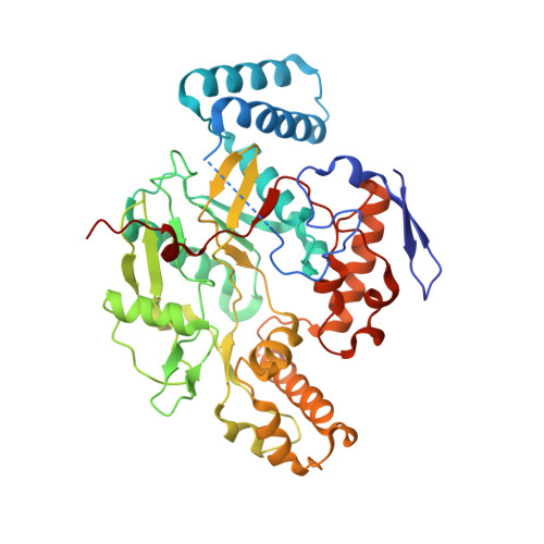
    The X-ray structures of neuronal nitric oxide synthase (nNOS) with N(omega)-hydroxy-l-arginine (l-NHA) and CO (or NO) bound have been determined at 1.91-2.2 A resolution. Microspectrophotometric techniques confirmed reduced redox state and the status of diatomic ligand complexes during X-ray diffraction data collection. The structure of nNOS-NHA-NO, a close mimic to the dioxygen complex, provides a picture of the potential interactions between the heme-bound diatomic ligand, substrate l-NHA, and the surrounding protein and solvent structure environment. The OH group of l-NHA in the X-ray structures deviates from the plane of the guanidinium moiety substantially, indicating that the OH-bearing, protonated guanidine N(omega) nitrogen of l-NHA has substantial sp(3) hybridization character. This nitrogen geometry, different from that of the guanidinium N(omega) nitrogen of l-arginine, allows a hydrogen bond to be donated to the proximal oxygen of the heme-bound dioxygen complex, thus preventing cleavage of the O-O bond. Instead, it favors the stabilization of the ferric-hydroperoxy intermediate, Fe(3+)-OOH(-), which serves as the active oxidant in the conversion of l-NHA to NO and citrulline in the second reaction of the NOS.


  • Organizational Affiliation

    Macromolecular Crystallographic Group, The Stanford Synchrotron Radiation Lightsource, SLAC, Stanford University, Stanford, California 94309, USA.


Macromolecules
Find similar proteins by:  (by identity cutoff)  |  3D Structure
Entity ID: 1
MoleculeChains Sequence LengthOrganismDetailsImage
Nitric oxide synthase, brain
A, B
422Rattus norvegicusMutation(s): 0 
Gene Names: Nos1Bnos
EC: 1.14.13.39
UniProt
Find proteins for P29476 (Rattus norvegicus)
Explore P29476 
Go to UniProtKB:  P29476
Entity Groups  
Sequence Clusters30% Identity50% Identity70% Identity90% Identity95% Identity100% Identity
UniProt GroupP29476
Sequence Annotations
Expand
  • Reference Sequence
Small Molecules
Ligands 7 Unique
IDChains Name / Formula / InChI Key2D Diagram3D Interactions
HEM
Query on HEM

Download Ideal Coordinates CCD File 
C [auth A],
K [auth B]
PROTOPORPHYRIN IX CONTAINING FE
C34 H32 Fe N4 O4
KABFMIBPWCXCRK-RGGAHWMASA-L
H4B
Query on H4B

Download Ideal Coordinates CCD File 
E [auth A],
M [auth B]
5,6,7,8-TETRAHYDROBIOPTERIN
C9 H15 N5 O3
FNKQXYHWGSIFBK-RPDRRWSUSA-N
HAR
Query on HAR

Download Ideal Coordinates CCD File 
F [auth A],
N [auth B]
N-OMEGA-HYDROXY-L-ARGININE
C6 H14 N4 O3
FQWRAVYMZULPNK-BYPYZUCNSA-N
GOL
Query on GOL

Download Ideal Coordinates CCD File 
I [auth A],
J [auth A],
P [auth B],
Q [auth B],
R [auth B]
GLYCEROL
C3 H8 O3
PEDCQBHIVMGVHV-UHFFFAOYSA-N
ZN
Query on ZN

Download Ideal Coordinates CCD File 
H [auth A]ZINC ION
Zn
PTFCDOFLOPIGGS-UHFFFAOYSA-N
ACT
Query on ACT

Download Ideal Coordinates CCD File 
G [auth A],
O [auth B]
ACETATE ION
C2 H3 O2
QTBSBXVTEAMEQO-UHFFFAOYSA-M
NO
Query on NO

Download Ideal Coordinates CCD File 
D [auth A],
L [auth B]
NITRIC OXIDE
N O
ODUCDPQEXGNKDN-UHFFFAOYSA-N
Experimental Data & Validation

Experimental Data

  • Method: X-RAY DIFFRACTION
  • Resolution: 2.20 Å
  • R-Value Free: 0.217 
  • R-Value Work: 0.164 
  • R-Value Observed: 0.167 
  • Space Group: P 21 21 21
Unit Cell:
Length ( Å )Angle ( ˚ )
a = 51.82α = 90
b = 110.56β = 90
c = 164.53γ = 90
Software Package:
Software NamePurpose
PHENIXrefinement
REFMACrefinement
Blu-Icedata collection
XDSdata reduction
XDSdata scaling
REFMACphasing

Structure Validation

View Full Validation Report


Ligand Structure Quality Assessment 


Entry History 

Deposition Data

Revision History  (Full details and data files)

  • Version 1.0: 2009-10-20
    Type: Initial release
  • Version 1.1: 2011-07-13
    Changes: Non-polymer description, Version format compliance
  • Version 1.2: 2019-07-24
    Changes: Data collection, Refinement description
  • Version 1.3: 2023-09-06
    Changes: Data collection, Database references, Derived calculations, Refinement description