


Pterin-binding site mutation Y53A, N55A or F123A and activity of E. coli HPPK
Li, Y., Blaszczyk, J., Ji, X., Yan, H.To be published.
Experimental Data Snapshot
Starting Model: experimental
View more details
Entity ID: 1 | |||||
|---|---|---|---|---|---|
| Molecule | Chains | Sequence Length | Organism | Details | Image |
| HPPK | 158 | Escherichia coli K-12 | Mutation(s): 1 Gene Names: b0142, foIK, folK, JW0138 EC: 2.7.6.3 | 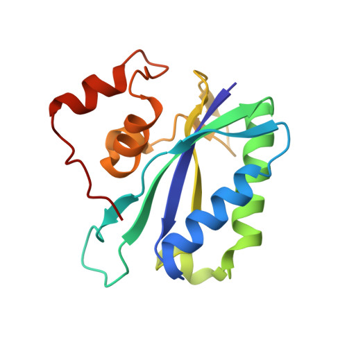 | |
UniProt | |||||
Entity Groups | |||||
| Sequence Clusters | 30% Identity50% Identity70% Identity90% Identity95% Identity100% Identity | ||||
| UniProt Group | P26281 | ||||
Sequence AnnotationsExpand | |||||
Reference Sequence | |||||
| Ligands 5 Unique | |||||
|---|---|---|---|---|---|
| ID | Chains | Name / Formula / InChI Key | 2D Diagram | 3D Interactions | |
| APC Download:Ideal Coordinates CCD File | D [auth A] | DIPHOSPHOMETHYLPHOSPHONIC ACID ADENOSYL ESTER C11 H18 N5 O12 P3 CAWZRIXWFRFUQB-IOSLPCCCSA-N |  | ||
| GOL Download:Ideal Coordinates CCD File | H [auth A], I [auth A] | GLYCEROL C3 H8 O3 PEDCQBHIVMGVHV-UHFFFAOYSA-N |  | ||
| ACT Download:Ideal Coordinates CCD File | F [auth A], G [auth A] | ACETATE ION C2 H3 O2 QTBSBXVTEAMEQO-UHFFFAOYSA-M |  | ||
| CL Download:Ideal Coordinates CCD File | E [auth A] | CHLORIDE ION Cl VEXZGXHMUGYJMC-UHFFFAOYSA-M |  | ||
| MG Download:Ideal Coordinates CCD File | B [auth A], C [auth A] | MAGNESIUM ION Mg JLVVSXFLKOJNIY-UHFFFAOYSA-N |  | ||
| Length ( Å ) | Angle ( ˚ ) |
|---|---|
| a = 35.94 | α = 90 |
| b = 48.02 | β = 90 |
| c = 74.2 | γ = 90 |
| Software Name | Purpose |
|---|---|
| DENZO | data reduction |
| SCALEPACK | data scaling |
| PHENIX | refinement |
| PDB_EXTRACT | data extraction |
| ADSC | data collection |
| AMoRE | phasing |


