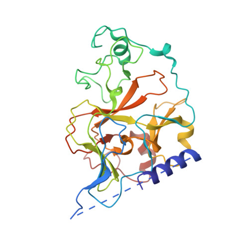
Structural biology of human H3K9 methyltransferases
Wu, H., Min, J., Lunin, V.V., Antoshenko, T., Dombrovski, L., Zeng, H., Allali-Hassani, A., Campagna-Slater, V., Vedadi, M., Arrowsmith, C.H., Plotnikov, A.N., Schapira, M.(2010) PLoS One 5: e8570-e8570
- PubMed: 20084102 Search on PubMedSearch on PubMed Central
- DOI: https://doi.org/10.1371/journal.pone.0008570
- Primary Citation Related Structures:
2IGQ, 2O8J, 2QPW, 2R3A, 2RFI, 3HNA - PubMed Abstract:
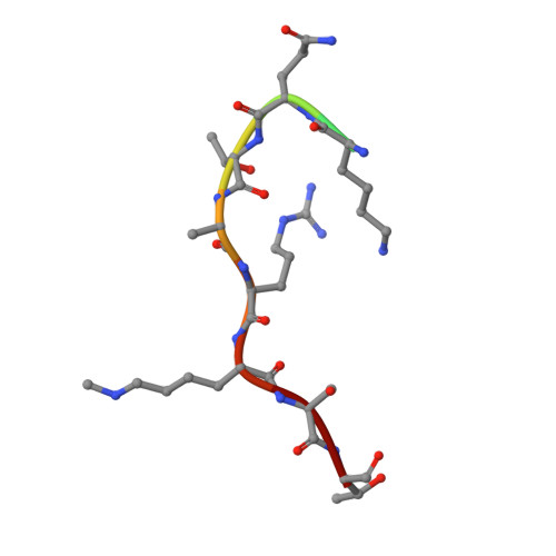
SET domain methyltransferases deposit methyl marks on specific histone tail lysine residues and play a major role in epigenetic regulation of gene transcription. We solved the structures of the catalytic domains of GLP, G9a, Suv39H2 and PRDM2, four of the eight known human H3K9 methyltransferases in their apo conformation or in complex with the methyl donating cofactor, and peptide substrates. We analyzed the structural determinants for methylation state specificity, and designed a G9a mutant able to tri-methylate H3K9. We show that the I-SET domain acts as a rigid docking platform, while induced-fit of the Post-SET domain is necessary to achieve a catalytically competent conformation. We also propose a model where long-range electrostatics bring enzyme and histone substrate together, while the presence of an arginine upstream of the target lysine is critical for binding and specificity. This article can also be viewed as an enhanced version in which the text of the article is integrated with interactive 3D representations and animated transitions. Please note that a web plugin is required to access this enhanced functionality. Instructions for the installation and use of the web plugin are available in Text S1.
- Structural Genomics Consortium, University of Toronto, Toronto, Ontario, Canada.
Organizational Affiliation: