3HMX | pdb_00003hmx

Crystal structure of ustekinumab FAB/IL-12 complex


Experimental Data Snapshot

  • Method: X-RAY DIFFRACTION
  • Resolution: 3.00 Å
  • R-Value Free: 
    0.303 (Depositor), 0.299 (DCC) 
  • R-Value Work: 
    0.211 (Depositor), 0.206 (DCC) 
  • R-Value Observed: 
    0.215 (Depositor) 

wwPDB Validation 3D Report Full Report

Validation slider image for 3HMX

This is version 2.1 of the entry. See complete history

Literature

Structural basis for the dual recognition of IL-12 and IL-23 by ustekinumab.

Luo, J.Wu, S.J.Lacy, E.R.Orlovsky, Y.Baker, A.Teplyakov, A.Obmolova, G.Heavner, G.A.Richter, H.T.Benson, J.

(2010) J Mol Biology 402: 797-812

  • DOI: https://doi.org/10.1016/j.jmb.2010.07.046
  • Primary Citation Related Structures: 
    3HMW, 3HMX

  • PubMed Abstract: 

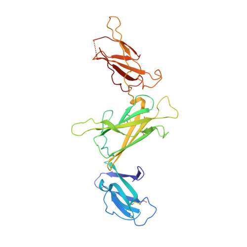
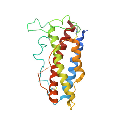
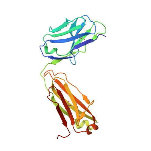
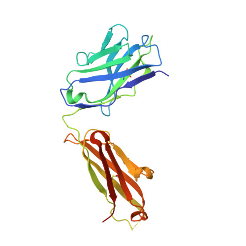
    Interleukin (IL)-12 and IL-23 are heterodimeric proinflammatory cytokines that share a common p40 subunit, paired with p35 and p19 subunits, respectively. They represent an attractive class of therapeutic targets for the treatment of psoriasis and other immune-mediated diseases. Ustekinumab is a fully human monoclonal antibody (mAb) that binds specifically to IL-12/IL-23p40 and neutralizes human IL-12 and IL-23 bioactivity. The crystal structure of ustekinumab Fab (antigen binding fragment of mAb), in complex with human IL-12, has been determined by X-ray crystallography at 3.0 Å resolution. Ustekinumab Fab binds the D1 domain of the p40 subunit in a 1:1 ratio in the crystal, consistent with a 2 cytokines:1 mAb stoichiometry, as measured by isothermal titration calorimetry. The structure indicates that ustekinumab binds to the same epitope on p40 in both IL-12 and IL-23 with identical interactions. Mutational analyses confirm that several residues identified in the IL-12/IL-23p40 epitope provide important molecular binding interactions with ustekinumab. The electrostatic complementarity between the mAb antigen binding site and the p40 D1 domain epitope appears to play a key role in antibody/antigen recognition specificity. Interestingly, this structure also reveals significant structural differences in the p35 subunit and p35/p40 interface, compared with the published crystal structure of human IL-12, suggesting unusual and potentially functionally relevant structural flexibility of p35, as well as p40/p35 recognition. Collectively, these data describe unique observations about IL-12p35 and ustekinumab interactions with p40 that account for its dual binding and neutralization of IL-12 and IL-23.


  • Organizational Affiliation
    • Centocor Research and Development, Inc., 145 King of Prussia Road, Radnor, PA 19087, USA. jluo@its.jnj.com

Macromolecule Content 

  • Total Structure Weight: 106.06 kDa 
  • Atom Count: 7,078 
  • Modeled Residue Count: 902 
  • Deposited Residue Count: 943 
  • Unique protein chains: 4

Macromolecules

Find similar proteins by:|  3D Structure
Entity ID: 1
MoleculeChains  Sequence LengthOrganismDetailsImage
Interleukin-12 subunit beta306Homo sapiensMutation(s): 0 
Gene Names: IL12BNKSF2
UniProt & NIH Common Fund Data Resources
Find proteins for P29460 (Homo sapiens)
Explore P29460 
Go to UniProtKB:  P29460
PHAROS:  P29460
GTEx:  ENSG00000113302 
Entity Groups
Sequence Clusters30% Identity50% Identity70% Identity90% Identity95% Identity100% Identity
UniProt GroupP29460
Glycosylation
Glycosylation Sites: 1Go to GlyGen: P29460-1
Sequence Annotations
Expand
Reference Sequence
Find similar proteins by:|  3D Structure
Entity ID: 2
MoleculeChains  Sequence LengthOrganismDetailsImage
Interleukin-12 subunit alpha197Homo sapiensMutation(s): 0 
Gene Names: IL12ANKSF1
UniProt & NIH Common Fund Data Resources
Find proteins for P29459 (Homo sapiens)
Explore P29459 
Go to UniProtKB:  P29459
PHAROS:  P29459
GTEx:  ENSG00000168811 
Entity Groups
Sequence Clusters30% Identity50% Identity70% Identity90% Identity95% Identity100% Identity
UniProt GroupP29459
Sequence Annotations
Expand
Reference Sequence
Find similar proteins by:|  3D Structure
Entity ID: 3
MoleculeChains  Sequence LengthOrganismDetailsImage
USTEKINUMAB FAB LIGHT CHAINC [auth L]214Homo sapiensMutation(s): 0 
Find similar proteins by:|  3D Structure
Entity ID: 4
MoleculeChains  Sequence LengthOrganismDetailsImage
USTEKINUMAB FAB HEAVY CHAIND [auth H]226Homo sapiensMutation(s): 0 

Oligosaccharides

Help  
Entity ID: 5
MoleculeChains Length2D Diagram GlycosylationD Interactions
alpha-D-mannopyranose-(1-3)-[alpha-D-mannopyranose-(1-6)]beta-D-mannopyranose-(1-4)-2-acetamido-2-deoxy-beta-D-glucopyranose-(1-4)-2-acetamido-2-deoxy-beta-D-glucopyranoseE [auth C]5N-Glycosylation
Glycosylation Resources
GlyTouCan: G22768VO
GlyCosmos: G22768VO
GlyGen: G22768VO

Experimental Data & Validation

Experimental Data

  • Method: X-RAY DIFFRACTION
  • Resolution: 3.00 Å
  • R-Value Free:  0.303 (Depositor), 0.299 (DCC) 
  • R-Value Work:  0.211 (Depositor), 0.206 (DCC) 
  • R-Value Observed: 0.215 (Depositor) 
Space Group: P 21 21 21
Unit Cell:
Length ( Å )Angle ( ˚ )
a = 55.092α = 90
b = 116.435β = 90
c = 182.781γ = 90
Software Package:
Software NamePurpose
d*TREKdata processing
PHENIXrefinement
PDB_EXTRACTdata extraction
ADSCdata collection
d*TREKdata reduction
d*TREKdata scaling
CNXphasing

Structure Validation

View Full Validation Report


Entry History 

Deposition Data

  • Released Date: 2010-06-09 
  • Deposition Author(s): Luo, J.

Revision History  (Full details and data files)

  • Version 1.0: 2010-06-09
    Type: Initial release
  • Version 1.1: 2011-07-13
    Changes: Version format compliance
  • Version 1.2: 2015-07-22
    Changes: Database references
  • Version 2.0: 2020-07-29
    Type: Remediation
    Reason: Carbohydrate remediation
    Changes: Advisory, Atomic model, Data collection, Derived calculations, Structure summary
  • Version 2.1: 2024-11-27
    Changes: Data collection, Database references, Structure summary