3HKL | pdb_00003hkl

Crystal Structure of the Frizzled-like Cysteine-rich Domain of MuSK


Experimental Data Snapshot

  • Method: X-RAY DIFFRACTION
  • Resolution: 2.10 Å
  • R-Value Free: 
    0.264 (Depositor), 0.263 (DCC) 
  • R-Value Work: 
    0.226 (Depositor), 0.225 (DCC) 
  • R-Value Observed: 
    0.228 (Depositor) 

wwPDB Validation   3D Report Full Report


This is version 2.1 of the entry. See complete history


Literature

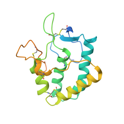
Crystal structure of the frizzled-like cysteine-rich domain of the receptor tyrosine kinase MuSK.

Stiegler, A.L.Burden, S.J.Hubbard, S.R.

(2009) J Mol Biology 393: 1-9

  • DOI: https://doi.org/10.1016/j.jmb.2009.07.091
  • Primary Citation of Related Structures:  
    3HKL

  • PubMed Abstract: 

    Muscle-specific kinase (MuSK) is an essential receptor tyrosine kinase for the establishment and maintenance of the neuromuscular junction (NMJ). Activation of MuSK by agrin, a neuronally derived heparan-sulfate proteoglycan, and LRP4 (low-density lipoprotein receptor-related protein-4), the agrin receptor, leads to clustering of acetylcholine receptors on the postsynaptic side of the NMJ. The ectodomain of MuSK comprises three immunoglobulin-like domains and a cysteine-rich domain (Fz-CRD) related to those in Frizzled proteins, the receptors for Wnts. Here, we report the crystal structure of the MuSK Fz-CRD at 2.1 A resolution. The structure reveals a five-disulfide-bridged domain similar to CRDs of Frizzled proteins but with a divergent C-terminal region. An asymmetric dimer present in the crystal structure implicates surface hydrophobic residues that may function in homotypic or heterotypic interactions to mediate co-clustering of MuSK, rapsyn, and acetylcholine receptors at the NMJ.


  • Organizational Affiliation
    • Structural Biology Program, Kimmel Center for Biology and Medicine of the Skirball Institute, New York University School of Medicine, New York, NY 10016, USA.

Macromolecules
Find similar proteins by:  (by identity cutoff)  |  3D Structure
Entity ID: 1
MoleculeChains Sequence LengthOrganismDetailsImage
Muscle, skeletal receptor tyrosine protein kinase
A, B
197Rattus norvegicusMutation(s): 0 
Gene Names: Musk
EC: 2.7.10.1
UniProt
Find proteins for Q62838 (Rattus norvegicus)
Explore Q62838 
Go to UniProtKB:  Q62838
Entity Groups  
Sequence Clusters30% Identity50% Identity70% Identity90% Identity95% Identity100% Identity
UniProt GroupQ62838
Glycosylation
Glycosylation Sites: 1Go to GlyGen: Q62838-1
Sequence Annotations
Expand
  • Reference Sequence
Oligosaccharides

Help

Entity ID: 2
MoleculeChains Length2D Diagram Glycosylation3D Interactions
2-acetamido-2-deoxy-beta-D-glucopyranose-(1-4)-2-acetamido-2-deoxy-beta-D-glucopyranose
C, D
2N-Glycosylation
Glycosylation Resources
GlyTouCan:  G42666HT
GlyCosmos:  G42666HT
GlyGen:  G42666HT
Experimental Data & Validation

Experimental Data

  • Method: X-RAY DIFFRACTION
  • Resolution: 2.10 Å
  • R-Value Free:  0.264 (Depositor), 0.263 (DCC) 
  • R-Value Work:  0.226 (Depositor), 0.225 (DCC) 
  • R-Value Observed: 0.228 (Depositor) 
Space Group: P 1 21 1
Unit Cell:
Length ( Å )Angle ( ˚ )
a = 65.592α = 90
b = 44.422β = 108.27
c = 69.53γ = 90
Software Package:
Software NamePurpose
REFMACrefinement
HKL-2000data collection
HKL-2000data reduction
HKL-2000data scaling

Structure Validation

View Full Validation Report


Entry History 

Deposition Data

Revision History  (Full details and data files)

  • Version 1.0: 2009-08-25
    Type: Initial release
  • Version 1.1: 2011-07-13
    Changes: Non-polymer description, Version format compliance
  • Version 2.0: 2020-07-29
    Type: Remediation
    Reason: Carbohydrate remediation
    Changes: Atomic model, Data collection, Database references, Derived calculations, Structure summary
  • Version 2.1: 2024-10-30
    Changes: Data collection, Database references, Structure summary