Mechanism-based tuning of a LOV domain photoreceptor.
Zoltowski, B.D., Vaccaro, B., Crane, B.R.(2009) Nat Chem Biol 5: 827-834
- PubMed: 19718042 Search on PubMedSearch on PubMed Central
- DOI: https://doi.org/10.1038/nchembio.210
- Primary Citation Related Structures:
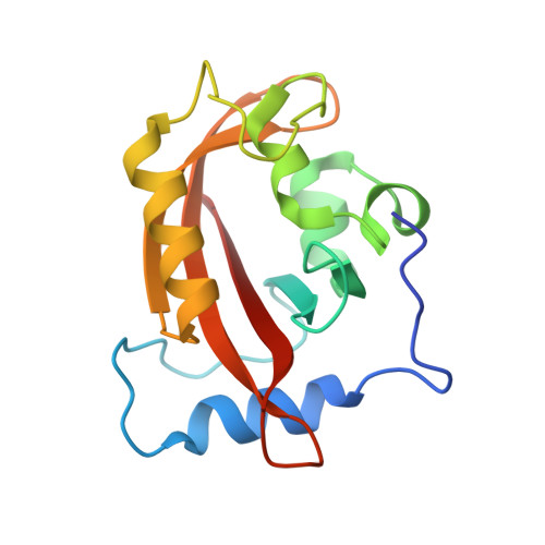
3HJI, 3HJK - PubMed Abstract:
Phototropin-like LOV domains form a cysteinyl-flavin adduct in response to blue light but show considerable variation in output signal and the lifetime of the photo-adduct signaling state. Mechanistic studies of the slow-cycling fungal LOV photoreceptor Vivid (VVD) reveal the importance of reactive cysteine conformation, flavin electronic environment and solvent accessibility for adduct scission and thermal reversion. Proton inventory, pH effects, base catalysis and structural studies implicate flavin N(5) deprotonation as rate-determining for recovery. Substitutions of active site residues Ile74, Ile85, Met135 and Met165 alter photoadduct lifetimes by over four orders of magnitude in VVD, and similar changes in other LOV proteins show analogous effects. Adduct state decay rates also correlate with changes in conformational and oligomeric properties of the protein necessary for signaling. These findings link natural sequence variation of LOV domains to function and provide a means to design broadly reactive light-sensitive probes.
- Department of Chemistry and Chemical Biology, Cornell University, Ithaca, New York, USA.
Organizational Affiliation: