3HIA | pdb_00003hia

Crystal structure of the choline binding domain of Spr1274 in Streptococcus pneumoniae


Experimental Data Snapshot

  • Method: X-RAY DIFFRACTION
  • Resolution: 2.38 Å
  • R-Value Free: 
    0.265 (Depositor), 0.273 (DCC) 
  • R-Value Work: 
    0.223 (Depositor), 0.236 (DCC) 
  • R-Value Observed: 
    0.229 (Depositor) 

Starting Model: experimental
View more details

wwPDB Validation 3D Report Full Report

Validation slider image for 3HIA

Ligand Structure Quality Assessment 


This is version 1.3 of the entry. See complete history

Literature

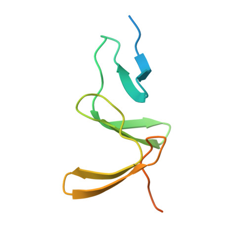
Structure of the choline-binding domain of Spr1274 in Streptococcus pneumoniae.

Zhang, Z.Li, W.Frolet, C.Bao, R.di Guilmi, A.M.Vernet, T.Chen, Y.

(2009) Acta Crystallogr Sect F Struct Biol Cryst Commun 65: 757-761

  • DOI: https://doi.org/10.1107/S1744309109025329
  • Primary Citation Related Structures: 
    3HIA

  • PubMed Abstract: 

    Spr1274 is a putative choline-binding protein that is bound to the cell wall of Streptococcus pneumoniae through noncovalent interactions with the choline moieties of teichoic and lipoteichoic acids. Its function is still unknown. The crystal structure of the choline-binding domain of Spr1274 (residues 44-129) was solved at 2.38 A resolution with three molecules in the asymmetric unit. It may provide a structural basis for functional analysis of choline-binding proteins.


  • Organizational Affiliation
    • Protein Research Institute, Tongji University, Shanghai 200092, People's Republic of China.

Macromolecule Content 

  • Total Structure Weight: 34.06 kDa 
  • Atom Count: 1,973 
  • Modeled Residue Count: 223 
  • Deposited Residue Count: 282 
  • Unique protein chains: 1

Macromolecules

Find similar proteins by:|  3D Structure
Entity ID: 1
MoleculeChains  Sequence LengthOrganismDetailsImage
Choline binding protein
A, B, C
94Streptococcus pneumoniae R6Mutation(s): 0 
Gene Names: spr1274
UniProt
Find proteins for Q8DP99 (Streptococcus pneumoniae (strain ATCC BAA-255 / R6))
Explore Q8DP99 
Go to UniProtKB:  Q8DP99
Entity Groups
Sequence Clusters30% Identity50% Identity70% Identity90% Identity95% Identity100% Identity
UniProt GroupQ8DP99
Sequence Annotations
Expand
Reference Sequence

Small Molecules

Ligands 2 Unique
IDChains Name / Formula / InChI Key2D Diagram3D Interactions
PC

Query on PC


Download:Ideal Coordinates CCD File
D [auth A]
F [auth B]
G [auth B]
H [auth B]
I [auth B]
D [auth A],
F [auth B],
G [auth B],
H [auth B],
I [auth B],
K [auth C],
L [auth C]
PHOSPHOCHOLINE
C5 H15 N O4 P
YHHSONZFOIEMCP-UHFFFAOYSA-O
SO4

Query on SO4


Download:Ideal Coordinates CCD File
E [auth A],
J [auth B],
M [auth C]
SULFATE ION
O4 S
QAOWNCQODCNURD-UHFFFAOYSA-L

Experimental Data & Validation

Experimental Data

  • Method: X-RAY DIFFRACTION
  • Resolution: 2.38 Å
  • R-Value Free:  0.265 (Depositor), 0.273 (DCC) 
  • R-Value Work:  0.223 (Depositor), 0.236 (DCC) 
  • R-Value Observed: 0.229 (Depositor) 
Space Group: H 3
Unit Cell:
Length ( Å )Angle ( ˚ )
a = 140.61α = 90
b = 140.61β = 90
c = 46.05γ = 120
Software Package:
Software NamePurpose
MAR345dtbdata collection
MOLREPphasing
REFMACrefinement
MOSFLMdata reduction
SCALEPACKdata scaling

Structure Validation

View Full Validation Report


Ligand Structure Quality Assessment 


Entry History 

Deposition Data

Revision History  (Full details and data files)

  • Version 1.0: 2009-08-04
    Type: Initial release
  • Version 1.1: 2011-07-13
    Changes: Source and taxonomy, Version format compliance
  • Version 1.2: 2023-11-01
    Changes: Data collection, Database references, Derived calculations, Refinement description
  • Version 1.3: 2025-05-07
    Changes: Database references, Structure summary