


Crystal structure of para-aminobenzoate synthetase, component I from Cytophaga hutchinsonii
Sampathkumar, P., Atwell, S., Wasserman, S., Do, J., Bain, K., Rutter, M., Gheyi, T., Sauder, J.M., Burley, S.K.To be published.
Experimental Data Snapshot
Starting Model: other
View more details
Entity ID: 1 | |||||
|---|---|---|---|---|---|
| Molecule | Chains | Sequence Length | Organism | Details | Image |
| p-aminobenzoate synthetase, component I | 436 | Cytophaga hutchinsonii ATCC 33406 | Mutation(s): 0 Gene Names: CHU_1808, pabB, YP_678417.1 EC: 4.1.3 | 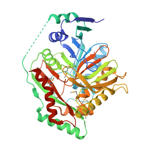 | |
| Ligands 2 Unique | |||||
|---|---|---|---|---|---|
| ID | Chains | Name / Formula / InChI Key | 2D Diagram | 3D Interactions | |
| DTU Download:Ideal Coordinates CCD File | B [auth A] | (2R,3S)-1,4-DIMERCAPTOBUTANE-2,3-DIOL C4 H10 O2 S2 VHJLVAABSRFDPM-ZXZARUISSA-N |  | ||
| PGE Download:Ideal Coordinates CCD File | C [auth A] | TRIETHYLENE GLYCOL C6 H14 O4 ZIBGPFATKBEMQZ-UHFFFAOYSA-N |  | ||
| Modified Residues 1 Unique | |||||
|---|---|---|---|---|---|
| ID | Chains | Type | Formula | 2D Diagram | Parent |
| MSE Query on MSE | A | L-PEPTIDE LINKING | C5 H11 N O2 Se |  | MET |
| Length ( Å ) | Angle ( ˚ ) |
|---|---|
| a = 60.006 | α = 90 |
| b = 159.497 | β = 90 |
| c = 115.542 | γ = 90 |
| Software Name | Purpose |
|---|---|
| REFMAC | refinement |
| PDB_EXTRACT | data extraction |
| ADSC | data collection |
| MOSFLM | data reduction |
| SCALA | data scaling |
| HKL2Map | phasing |


