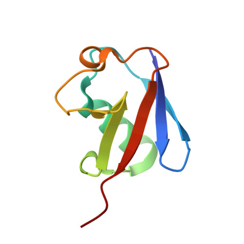
Crystal structures of Lys-63-linked tri- and di-ubiquitin reveal a highly extended chain architecture.
Weeks, S.D., Grasty, K.C., Hernandez-Cuebas, L., Loll, P.J.(2009) Proteins 77: 753-759
- PubMed: 19731378 Search on PubMedSearch on PubMed Central
- DOI: https://doi.org/10.1002/prot.22568
- Primary Citation Related Structures:
3H7P, 3H7S - PubMed Abstract:
The covalent attachment of different types of poly-ubiquitin chains signal different outcomes for the proteins so targeted. For example, a protein modified with Lys-48-linked poly-ubiquitin chains is targeted for proteasomal degradation, whereas Lys-63-linked chains encode nondegradative signals. The structural features that enable these different types of chains to encode different signals have not yet been fully elucidated. We report here the X-ray crystal structures of Lys-63-linked tri- and di-ubiquitin at resolutions of 2.3 and 1.9 A, respectively. The tri- and di-ubiquitin species adopt essentially identical structures. In both instances, the ubiquitin chain assumes a highly extended conformation with a left-handed helical twist; the helical chain contains four ubiquitin monomers per turn and has a repeat length of approximately 110 A. Interestingly, Lys-48 ubiquitin chains also adopt a left-handed helical structure with a similar repeat length. However, the Lys-63 architecture is much more open than that of Lys-48 chains and exposes much more of the ubiquitin surface for potential recognition events. These new crystal structures are consistent with the results of solution studies of Lys-63 chain conformation, and reveal the structural basis for differential recognition of Lys-63 versus Lys-48 chains.
- Department of Biochemistry and Molecular Biology, Drexel University College of Medicine, Philadelphia, Pennsylvania 19102, USA.
Organizational Affiliation: