


Crystal Structure of a Periplasmic Sugar-binding protein from the Pseudomonas fluorescens
Kumaran, D., Mahmood, A., Burley, S.K., Swaminathan, S.To be published.
Experimental Data Snapshot
wwPDB Validation 3D Report Full Report

Entity ID: 1 | |||||
|---|---|---|---|---|---|
| Molecule | Chains | Sequence Length | Organism | Details | Image |
| Periplasmic sugar-binding domain protein | 350 | Pseudomonas protegens Pf-5 | Mutation(s): 0 Gene Names: PFL_3664 | 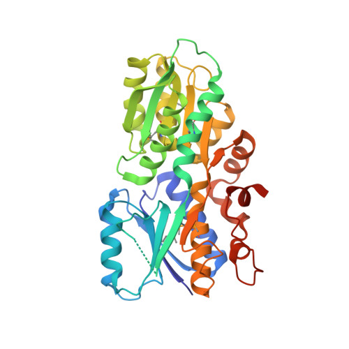 | |
UniProt | |||||
Entity Groups | |||||
| Sequence Clusters | 30% Identity50% Identity70% Identity90% Identity95% Identity100% Identity | ||||
| UniProt Group | Q4KAG7 | ||||
Sequence AnnotationsExpand | |||||
Reference Sequence | |||||
| Ligands 2 Unique | |||||
|---|---|---|---|---|---|
| ID | Chains | Name / Formula / InChI Key | 2D Diagram | 3D Interactions | |
| SO4 Download:Ideal Coordinates CCD File | B [auth A], C [auth A] | SULFATE ION O4 S QAOWNCQODCNURD-UHFFFAOYSA-L |  | ||
| GOL Download:Ideal Coordinates CCD File | D [auth A] | GLYCEROL C3 H8 O3 PEDCQBHIVMGVHV-UHFFFAOYSA-N |  | ||
| Modified Residues 1 Unique | |||||
|---|---|---|---|---|---|
| ID | Chains | Type | Formula | 2D Diagram | Parent |
| MSE Query on MSE | A | L-PEPTIDE LINKING | C5 H11 N O2 Se |  | MET |
| Length ( Å ) | Angle ( ˚ ) |
|---|---|
| a = 65.526 | α = 90 |
| b = 81.275 | β = 90 |
| c = 63.082 | γ = 90 |
| Software Name | Purpose |
|---|---|
| CBASS | data collection |
| SHELXD | phasing |
| SHELXE | model building |
| ARP/wARP | model building |
| CNS | refinement |
| HKL-2000 | data reduction |
| HKL-2000 | data scaling |


