3H6D

Structure of the mycobacterium tuberculosis DUTPase D28N mutant


Experimental Data Snapshot

  • Method: X-RAY DIFFRACTION
  • Resolution: 1.80 Å
  • R-Value Free: 0.207 
  • R-Value Work: 0.158 
  • R-Value Observed: 0.160 

wwPDB Validation   3D Report Full Report


Ligand Structure Quality Assessment 


This is version 1.3 of the entry. See complete history


Literature

Direct contacts between conserved motifs of different subunits provide major contribution to active site organization in human and mycobacterial dUTPases.

Takacs, E.Nagy, G.Leveles, I.Harmat, V.Lopata, A.Toth, J.Vertessy, B.G.

(2010) FEBS Lett 584: 3047-3054

  • DOI: https://doi.org/10.1016/j.febslet.2010.05.018
  • Primary Citation of Related Structures:  
    3H6D, 3I93

  • PubMed Abstract: 

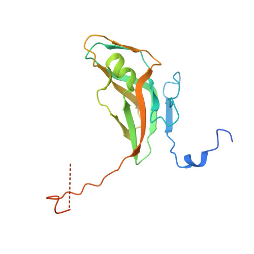
    dUTP pyrophosphatases (dUTPases) are essential for genome integrity. Recent results allowed characterization of the role of conserved residues. Here we analyzed the Asp/Asn mutation within conserved Motif I of human and mycobacterial dUTPases, wherein the Asp residue was previously implicated in Mg(2+)-coordination. Our results on transient/steady-state kinetics, ligand binding and a 1.80 A resolution structure of the mutant mycobacterial enzyme, in comparison with wild type and C-terminally truncated structures, argue that this residue has a major role in providing intra- and intersubunit contacts, but is not essential for Mg(2+) accommodation. We conclude that in addition to the role of conserved motifs in substrate accommodation, direct subunit interaction between protein atoms of active site residues from different conserved motifs are crucial for enzyme function.


  • Organizational Affiliation

    Institute of Enzymology, BRC, Hungarian Academy of Sciences, Budapest, Hungary.


Macromolecules
Find similar proteins by:  (by identity cutoff)  |  3D Structure
Entity ID: 1
MoleculeChains Sequence LengthOrganismDetailsImage
Deoxyuridine 5'-triphosphate nucleotidohydrolase174Mycobacterium tuberculosisMutation(s): 1 
Gene Names: dutMT2771MTCY05A6.18cRv2697c
EC: 3.6.1.23
UniProt
Find proteins for P9WNS5 (Mycobacterium tuberculosis (strain ATCC 25618 / H37Rv))
Explore P9WNS5 
Go to UniProtKB:  P9WNS5
Entity Groups  
Sequence Clusters30% Identity50% Identity70% Identity90% Identity95% Identity100% Identity
UniProt GroupP9WNS5
Sequence Annotations
Expand
  • Reference Sequence
Experimental Data & Validation

Experimental Data

  • Method: X-RAY DIFFRACTION
  • Resolution: 1.80 Å
  • R-Value Free: 0.207 
  • R-Value Work: 0.158 
  • R-Value Observed: 0.160 
  • Space Group: P 63
Unit Cell:
Length ( Å )Angle ( ˚ )
a = 55.228α = 90
b = 55.228β = 90
c = 83.65γ = 120
Software Package:
Software NamePurpose
XSCALEdata scaling
MOLREPphasing
REFMACrefinement
PDB_EXTRACTdata extraction
XDSdata reduction

Structure Validation

View Full Validation Report


Ligand Structure Quality Assessment 


Entry History 

Deposition Data

Revision History  (Full details and data files)

  • Version 1.0: 2009-11-24
    Type: Initial release
  • Version 1.1: 2011-07-13
    Changes: Refinement description, Version format compliance
  • Version 1.2: 2021-10-13
    Changes: Data collection, Database references, Derived calculations
  • Version 1.3: 2023-09-06
    Changes: Data collection, Refinement description