3H40 | pdb_00003h40

Binary complex of human DNA polymerase iota with template U/T


Experimental Data Snapshot

  • Method: X-RAY DIFFRACTION
  • Resolution: 2.30 Å
  • R-Value Free: 
    0.277 (Depositor), 0.278 (DCC) 
  • R-Value Work: 
    0.253 (Depositor), 0.247 (DCC) 
  • R-Value Observed: 
    0.253 (Depositor) 

Starting Model: experimental
View more details

wwPDB Validation 3D Report Full Report

Validation slider image for 3H40

This is version 1.2 of the entry. See complete history

Literature

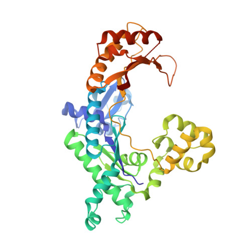
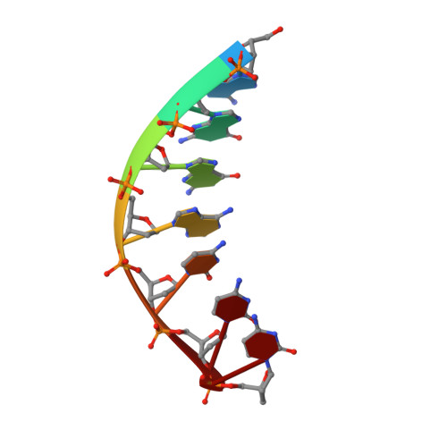
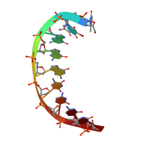
Replication across template T/U by human DNA polymerase-iota.

Jain, R.Nair, D.T.Johnson, R.E.Prakash, L.Prakash, S.Aggarwal, A.K.

(2009) Structure 17: 974-980

  • DOI: https://doi.org/10.1016/j.str.2009.04.011
  • Primary Citation Related Structures: 
    3H40, 3H4B, 3H4D

  • PubMed Abstract: 

    Human DNA polymerase-iota (Poliota) incorporates correct nucleotides opposite template purines with a much higher efficiency and fidelity than opposite template pyrimidines. In fact, the fidelity opposite template T is so poor that Poliota inserts an incorrect dGTP approximately 10 times better than it inserts the correct dATP. We determine here how a template T/U is accommodated in the Poliota active site and why a G is incorporated more efficiently than an A. We show that in the absence of incoming dATP or dGTP (binary complex), template T/U exists in both syn and anti conformations, but in the presence of dATP or dGTP (ternary complexes), template T/U is predominantly in the anti conformation. We also show that dATP and dGTP insert differently opposite template T/U, and that the basis of selection of dGTP over dATP is a hydrogen bond between the N2 amino group of dGTP and Gln59 of Poliota.


  • Organizational Affiliation
    • Department of Structural and Chemical Biology, Mount Sinai School of Medicine, New York, NY 10029, USA.

Macromolecule Content 

  • Total Structure Weight: 48.96 kDa 
  • Atom Count: 3,391 
  • Modeled Residue Count: 388 
  • Deposited Residue Count: 405 
  • Unique protein chains: 1
  • Unique nucleic acid chains: 2

Macromolecules


Find similar proteins by:|  3D Structure
Entity ID: 1
MoleculeChains  Sequence LengthOrganismDetailsImage
DNA polymerase iota389Homo sapiensMutation(s): 0 
Gene Names: POLIRAD30B
EC: 2.7.7.7
UniProt & NIH Common Fund Data Resources
Find proteins for Q9UNA4 (Homo sapiens)
Explore Q9UNA4 
Go to UniProtKB:  Q9UNA4
PHAROS:  Q9UNA4
GTEx:  ENSG00000101751 
Entity Groups
Sequence Clusters30% Identity50% Identity70% Identity90% Identity95% Identity100% Identity
UniProt GroupQ9UNA4
Sequence Annotations
Expand
Reference Sequence
Find similar nucleic acids by:  Sequence
Entity ID: 2
MoleculeChains LengthOrganismImage
5'-D(*TP*(BRU)P*GP*GP*GP*TP*CP*CP*T)-3'B [auth P]7N/A
Sequence Annotations
Expand
Reference Sequence
Find similar nucleic acids by:  Sequence
Entity ID: 3
MoleculeChains LengthOrganismImage
5'-D(*AP*GP*GP*AP*CP*CP*(DOC))-3'C [auth T]9N/A
Sequence Annotations
Expand
Reference Sequence

Small Molecules

Ligands 1 Unique
IDChains Name / Formula / InChI Key2D Diagram3D Interactions
SO4

Query on SO4


Download:Ideal Coordinates CCD File
D [auth A]SULFATE ION
O4 S
QAOWNCQODCNURD-UHFFFAOYSA-L

Experimental Data & Validation

Experimental Data

  • Method: X-RAY DIFFRACTION
  • Resolution: 2.30 Å
  • R-Value Free:  0.277 (Depositor), 0.278 (DCC) 
  • R-Value Work:  0.253 (Depositor), 0.247 (DCC) 
  • R-Value Observed: 0.253 (Depositor) 
Space Group: P 65 2 2
Unit Cell:
Length ( Å )Angle ( ˚ )
a = 98.243α = 90
b = 98.243β = 90
c = 203.528γ = 120
Software Package:
Software NamePurpose
ADSCdata collection
AMoREphasing
CNSrefinement
HKL-2000data reduction
HKL-2000data scaling

Structure Validation

View Full Validation Report


Entry History 

Deposition Data

Revision History  (Full details and data files)

  • Version 1.0: 2009-07-21
    Type: Initial release
  • Version 1.1: 2011-07-13
    Changes: Version format compliance
  • Version 1.2: 2023-09-06
    Changes: Data collection, Database references, Derived calculations, Refinement description