3H2A | pdb_00003h2a

Structural Studies of Pterin-Based Inhibitors of Dihydropteroate Synthase


Experimental Data Snapshot

  • Method: X-RAY DIFFRACTION
  • Resolution: 2.40 Å
  • R-Value Free: 
    0.280 (Depositor), 0.276 (DCC) 
  • R-Value Work: 
    0.225 (Depositor), 0.221 (DCC) 
  • R-Value Observed: 
    0.228 (Depositor) 

Starting Model: experimental
View more details

wwPDB Validation 3D Report Full Report

Validation slider image for 3H2A

Ligand Structure Quality Assessment 


This is version 1.3 of the entry. See complete history

Literature

Structural studies of pterin-based inhibitors of dihydropteroate synthase.

Hevener, K.E.Yun, M.K.Qi, J.Kerr, I.D.Babaoglu, K.Hurdle, J.G.Balakrishna, K.White, S.W.Lee, R.E.

(2010) J Med Chem 53: 166-177

  • DOI: https://doi.org/10.1021/jm900861d
  • Primary Citation Related Structures: 
    3H21, 3H22, 3H23, 3H24, 3H26, 3H2A, 3H2C, 3H2E, 3H2F, 3H2M, 3H2N, 3H2O

  • PubMed Abstract: 

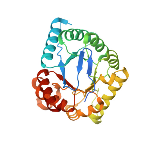
    Dihydropteroate synthase (DHPS) is a key enzyme in bacterial folate synthesis and the target of the sulfonamide class of antibacterials. Resistance and toxicities associated with sulfonamides have led to a decrease in their clinical use. Compounds that bind to the pterin binding site of DHPS, as opposed to the p-amino benzoic acid (pABA) binding site targeted by the sulfonamide agents, are anticipated to bypass sulfonamide resistance. To identify such inhibitors and map the pterin binding pocket, we have performed virtual screening, synthetic, and structural studies using Bacillus anthracis DHPS. Several compounds with inhibitory activity have been identified, and crystal structures have been determined that show how the compounds engage the pterin site. The structural studies identify the key binding elements and have been used to generate a structure-activity based pharmacophore map that will facilitate the development of the next generation of DHPS inhibitors which specifically target the pterin site.


  • Organizational Affiliation
    • Department of Pharmaceutical Sciences, University of Tennessee Health Science Center, 847 Monroe Avenue, Room 327 Johnson Building, Memphis, Tennessee 38163, USA.

Macromolecule Content 

  • Total Structure Weight: 66.78 kDa 
  • Atom Count: 4,333 
  • Modeled Residue Count: 538 
  • Deposited Residue Count: 594 
  • Unique protein chains: 1

Macromolecules

Find similar proteins by:|  3D Structure
Entity ID: 1
MoleculeChains  Sequence LengthOrganismDetailsImage
Dihydropteroate synthase
A, B
297Bacillus anthracis str. A2012Mutation(s): 0 
Gene Names: folPBAO_0074
EC: 2.5.1.15
UniProt
Find proteins for Q81VW8 (Bacillus anthracis)
Explore Q81VW8 
Go to UniProtKB:  Q81VW8
Entity Groups
Sequence Clusters30% Identity50% Identity70% Identity90% Identity95% Identity100% Identity
UniProt GroupQ81VW8
Sequence Annotations
Expand
Reference Sequence

Small Molecules

Ligands 2 Unique
IDChains Name / Formula / InChI Key2D Diagram3D Interactions
B57

Query on B57


Download:Ideal Coordinates CCD File
C [auth A],
G [auth B]
2,6-diamino-5-nitropyrimidin-4(3H)-one
C4 H5 N5 O3
XNFGVBWYGFPSDN-UHFFFAOYSA-N
SO4

Query on SO4


Download:Ideal Coordinates CCD File
D [auth A]
E [auth A]
F [auth A]
H [auth B]
I [auth B]
D [auth A],
E [auth A],
F [auth A],
H [auth B],
I [auth B],
J [auth B],
K [auth B]
SULFATE ION
O4 S
QAOWNCQODCNURD-UHFFFAOYSA-L
Binding Affinity Annotations 
IDSourceBinding Affinity
B57 BindingDB:  3H2A IC50: 1.09e+5 (nM) from 1 assay(s)

Experimental Data & Validation

Experimental Data

  • Method: X-RAY DIFFRACTION
  • Resolution: 2.40 Å
  • R-Value Free:  0.280 (Depositor), 0.276 (DCC) 
  • R-Value Work:  0.225 (Depositor), 0.221 (DCC) 
  • R-Value Observed: 0.228 (Depositor) 
Space Group: P 62 2 2
Unit Cell:
Length ( Å )Angle ( ˚ )
a = 97.513α = 90
b = 97.513β = 90
c = 261.643γ = 120
Software Package:
Software NamePurpose
SERGUIdata collection
REFMACrefinement
HKL-2000data reduction
HKL-2000data scaling

Structure Validation

View Full Validation Report


Ligand Structure Quality Assessment 


Entry History 

Deposition Data

Revision History  (Full details and data files)

  • Version 1.0: 2009-12-08
    Type: Initial release
  • Version 1.1: 2011-07-13
    Changes: Advisory, Version format compliance
  • Version 1.2: 2017-11-01
    Changes: Refinement description
  • Version 1.3: 2023-09-06
    Changes: Data collection, Database references, Derived calculations, Refinement description