3H1V | pdb_00003h1v

Human glucokinase in complex with a synthetic activator


Experimental Data Snapshot

  • Method: X-RAY DIFFRACTION
  • Resolution: 2.11 Å
  • R-Value Free: 
    0.284 (Depositor), 0.271 (DCC) 
  • R-Value Work: 
    0.232 (Depositor) 
  • R-Value Observed: 
    0.235 (Depositor) 

wwPDB Validation 3D Report Full Report

Validation slider image for 3H1V

Ligand Structure Quality Assessment 


This is version 1.4 of the entry. See complete history

Literature

The design and optimization of a series of 2-(pyridin-2-yl)-1H-benzimidazole compounds as allosteric glucokinase activators.

Takahashi, K.Hashimoto, N.Nakama, C.Kamata, K.Sasaki, K.Yoshimoto, R.Ohyama, S.Hosaka, H.Maruki, H.Nagata, Y.Eiki, J.Nishimura, T.

(2009) Bioorg Med Chem 17: 7042-7051

  • DOI: https://doi.org/10.1016/j.bmc.2009.05.037
  • Primary Citation Related Structures: 
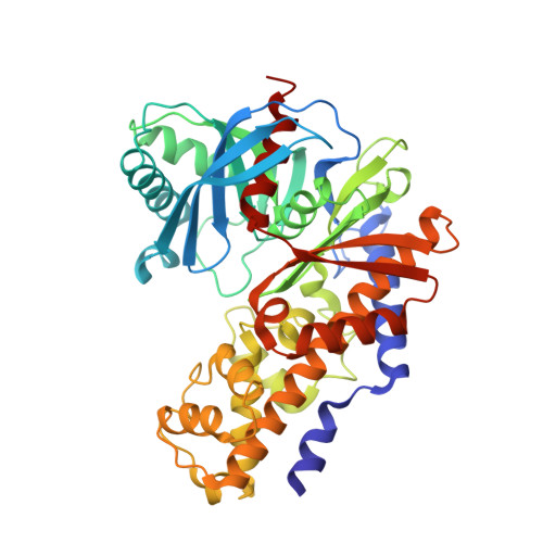
    3H1V

  • PubMed Abstract: 

    The optimization of a series of benzimidazole glucokinase activators is described. We identified a novel and potent achiral benzimidazole derivative as an allosteric GK activator. This activator was designed and synthesized via removal of the chiral center of the lead compound, 6-(N-acylpyrrolidin-2-yl)benzimidazole. The activator exhibited good PK profiles in rats and dogs, and significant hypoglycemic efficacy at 1 mg/kg po dosing in a rat OGTT model. The binding site and binding mode of the benzimidazole class of GKA with GK protein was confirmed by X-ray crystallographic analysis.


  • Organizational Affiliation
    • Banyu Tsukuba Research Institute, Banyu Pharmaceutical Co., Ltd, 3 Okubo, Tsukuba, Ibaraki 300-2611, Japan. g94e079@ruri.waseda.jp

Macromolecule Content 

  • Total Structure Weight: 51.29 kDa 
  • Atom Count: 3,659 
  • Modeled Residue Count: 446 
  • Deposited Residue Count: 451 
  • Unique protein chains: 1

Macromolecules

Find similar proteins by:|  3D Structure
Entity ID: 1
MoleculeChains  Sequence LengthOrganismDetailsImage
GlucokinaseA [auth X]451Homo sapiensMutation(s): 0 
Gene Names: GCK
EC: 2.7.1.2 (PDB Primary Data), 2.7.1.1 (UniProt)
UniProt & NIH Common Fund Data Resources
Find proteins for P35557 (Homo sapiens)
Explore P35557 
Go to UniProtKB:  P35557
PHAROS:  P35557
GTEx:  ENSG00000106633 
Entity Groups
Sequence Clusters30% Identity50% Identity70% Identity90% Identity95% Identity100% Identity
UniProt GroupP35557
Sequence Annotations
Expand
Reference Sequence

Small Molecules

Ligands 3 Unique
IDChains Name / Formula / InChI Key2D Diagram3D Interactions
TK1

Query on TK1


Download:Ideal Coordinates CCD File
C [auth X]1-({5-[4-(methylsulfonyl)phenoxy]-2-pyridin-2-yl-1H-benzimidazol-6-yl}methyl)pyrrolidine-2,5-dione
C24 H20 N4 O5 S
IPZLCOKXDRIZBC-UHFFFAOYSA-N
GLC

Query on GLC


Download:Ideal Coordinates CCD File
B [auth X]alpha-D-glucopyranose
C6 H12 O6
WQZGKKKJIJFFOK-DVKNGEFBSA-N
NA

Query on NA


Download:Ideal Coordinates CCD File
D [auth X]SODIUM ION
Na
FKNQFGJONOIPTF-UHFFFAOYSA-N

Experimental Data & Validation

Experimental Data

  • Method: X-RAY DIFFRACTION
  • Resolution: 2.11 Å
  • R-Value Free:  0.284 (Depositor), 0.271 (DCC) 
  • R-Value Work:  0.232 (Depositor) 
  • R-Value Observed: 0.235 (Depositor) 
Space Group: P 65 2 2
Unit Cell:
Length ( Å )Angle ( ˚ )
a = 79.793α = 90
b = 79.793β = 90
c = 326.124γ = 120
Software Package:
Software NamePurpose
DENZOdata reduction
SCALEPACKdata scaling
REFMACrefinement
PDB_EXTRACTdata extraction

Structure Validation

View Full Validation Report


Ligand Structure Quality Assessment 


Entry History 

Deposition Data

Revision History  (Full details and data files)

  • Version 1.0: 2009-10-06
    Type: Initial release
  • Version 1.1: 2011-07-13
    Changes: Version format compliance
  • Version 1.2: 2017-11-01
    Changes: Refinement description
  • Version 1.3: 2020-07-29
    Type: Remediation
    Reason: Carbohydrate remediation
    Changes: Data collection, Database references, Derived calculations, Structure summary
  • Version 1.4: 2024-03-20
    Changes: Data collection, Database references, Structure summary