3GXV

Three-dimensional structure of N-terminal domain of DnaB Helicase from Helicobacter pylori and its interactions with primase


Experimental Data Snapshot

  • Method: X-RAY DIFFRACTION
  • Resolution: 2.20 Å
  • R-Value Free: 0.278 
  • R-Value Work: 0.249 
  • R-Value Observed: 0.249 

wwPDB Validation   3D Report Full Report


This is version 1.2 of the entry. See complete history


Literature

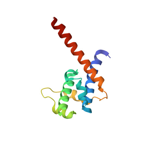
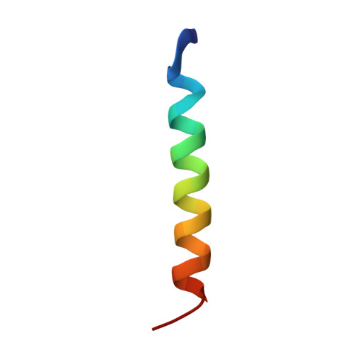
Three-dimensional structure of N-terminal domain of DnaB helicase and helicase-primase interactions in Helicobacter pylori

Kashav, T.Nitharwal, R.Abdulrehman, S.A.Gabdoulkhakov, A.Saenger, W.Dhar, S.K.Gourinath, S.

(2009) PLoS One 4: e7515-e7515

  • DOI: https://doi.org/10.1371/journal.pone.0007515
  • Primary Citation of Related Structures:  
    3GXV

  • PubMed Abstract: 

    Replication initiation is a crucial step in genome duplication and homohexameric DnaB helicase plays a central role in the replication initiation process by unwinding the duplex DNA and interacting with several other proteins during the process of replication. N-terminal domain of DnaB is critical for helicase activity and for DnaG primase interactions. We present here the crystal structure of the N-terminal domain (NTD) of H. pylori DnaB (HpDnaB) helicase at 2.2 A resolution and compare the structural differences among helicases and correlate with the functional differences. The structural details of NTD suggest that the linker region between NTD and C-terminal helicase domain plays a vital role in accurate assembly of NTD dimers. The sequence analysis of the linker regions from several helicases reveals that they should form four helix bundles. We also report the characterization of H. pylori DnaG primase and study the helicase-primase interactions, where HpDnaG primase stimulates DNA unwinding activity of HpDnaB suggesting presence of helicase-primase cohort at the replication fork. The protein-protein interaction study of C-terminal domain of primase and different deletion constructs of helicase suggests that linker is essential for proper conformation of NTD to interact strongly with HpDnaG. The surface charge distribution on the primase binding surface of NTDs of various helicases suggests that DnaB-DnaG interaction and stability of the complex is most probably charge dependent. Structure of the linker and helicase-primase interactions indicate that HpDnaB differs greatly from E.coli DnaB despite both belong to gram negative bacteria.


  • Organizational Affiliation

    School of Life Sciences, Jawaharlal Nehru University, New Delhi, India.


Macromolecules
Find similar proteins by:  (by identity cutoff)  |  3D Structure
Entity ID: 1
MoleculeChains Sequence LengthOrganismDetailsImage
Replicative DNA helicase
A, B
123Helicobacter pyloriMutation(s): 0 
Gene Names: HP_1362
EC: 3.6.1
UniProt
Find proteins for O25916 (Helicobacter pylori (strain ATCC 700392 / 26695))
Explore O25916 
Go to UniProtKB:  O25916
Entity Groups  
Sequence Clusters30% Identity50% Identity70% Identity90% Identity95% Identity100% Identity
UniProt GroupO25916
Sequence Annotations
Expand
  • Reference Sequence

Find similar proteins by:  Sequence   |   3D Structure  

Entity ID: 2
MoleculeChains Sequence LengthOrganismDetailsImage
Replicative DNA helicase22Helicobacter pyloriMutation(s): 0 
Gene Names: HP_1362
EC: 3.6.1
UniProt
Find proteins for O25916 (Helicobacter pylori (strain ATCC 700392 / 26695))
Explore O25916 
Go to UniProtKB:  O25916
Entity Groups  
Sequence Clusters30% Identity50% Identity70% Identity90% Identity95% Identity100% Identity
UniProt GroupO25916
Sequence Annotations
Expand
  • Reference Sequence
Find similar proteins by:  (by identity cutoff)  |  3D Structure
Entity ID: 3
MoleculeChains Sequence LengthOrganismDetailsImage
Replicative DNA helicase26Helicobacter pyloriMutation(s): 0 
Gene Names: HP_1362
EC: 3.6.1
UniProt
Find proteins for O25916 (Helicobacter pylori (strain ATCC 700392 / 26695))
Explore O25916 
Go to UniProtKB:  O25916
Entity Groups  
Sequence Clusters30% Identity50% Identity70% Identity90% Identity95% Identity100% Identity
UniProt GroupO25916
Sequence Annotations
Expand
  • Reference Sequence
Experimental Data & Validation

Experimental Data

  • Method: X-RAY DIFFRACTION
  • Resolution: 2.20 Å
  • R-Value Free: 0.278 
  • R-Value Work: 0.249 
  • R-Value Observed: 0.249 
  • Space Group: P 21 21 21
Unit Cell:
Length ( Å )Angle ( ˚ )
a = 71.83α = 90
b = 78.15β = 90
c = 108γ = 90
Software Package:
Software NamePurpose
HKL-2000data collection
PHASERphasing
CNSrefinement
AUTOMARdata reduction

Structure Validation

View Full Validation Report


Entry History 

Deposition Data

Revision History  (Full details and data files)

  • Version 1.0: 2010-01-26
    Type: Initial release
  • Version 1.1: 2011-07-13
    Changes: Version format compliance
  • Version 1.2: 2023-11-01
    Changes: Data collection, Database references, Refinement description